Bone remodeling and fracture repair
1Why this matters
Ruth, 79, trips while reaching into a kitchen cupboard and falls onto her side. The X-ray shows a broken hip, the upper end of her thigh bone. Her surgeon explains that a fall from standing would not break healthy bone: her bone had been thinning silently for thirty years, so an ordinary fall was enough to snap it. To understand how bone can quietly disappear, and how the same cells will now try to heal it, you need to know that adult bone is never finished.
2What this builds on
3Quick check before you start
1. What is the first event in the body's response to tissue injury, before repair begins?
- Scar tissue replaces the damaged cells
- Inflammation: vessels widen and leak, and the area swells
- Stem cells rebuild the tissue immediately
Show the answer
Injury triggers inflammation first: local vessels widen and become leaky, fluid and white blood cells enter, and macrophages begin clearing debris. Repair follows.
- Scar tissue replaces the damaged cells:
- Correct: Inflammation: vessels widen and leak, and the area swells:
- Stem cells rebuild the tissue immediately:
2. Which bone cell resorbs bone by dissolving mineral with acid and digesting collagen with enzymes?
- Osteoclast
- Osteoblast
- Osteogenic cell
Show the answer
Osteoclasts, huge fused cells of the macrophage line, pump out hydrogen ions and release lysosomal enzymes onto the bone.
- Correct: Osteoclast:
- Osteoblast:
- Osteogenic cell:
3. What forms when blood leaves a torn vessel and its proteins set into a mesh that traps blood cells?
- A callus
- A blood clot
- An abscess
Show the answer
Blood that escapes from a torn vessel clots. In a fracture, the clot around the break is the fracture hematoma, the first stage of repair.
- A callus:
- Correct: A blood clot:
- An abscess:
4Anatomy
With labels hidden, select a box to reveal its label.
5How it works, step by step
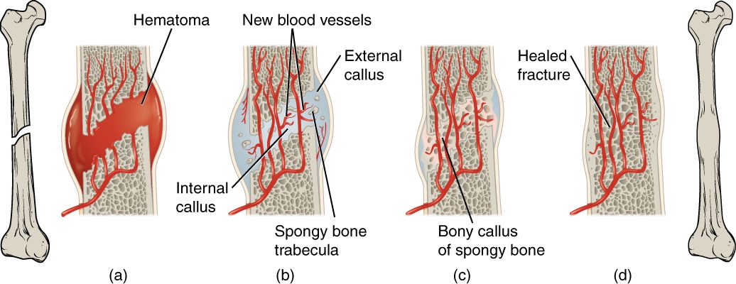
- A break tears blood vessels in the periosteum, bone and marrow.Blood pools and clots around the break, forming a fracture hematoma, and osteocytes at the broken ends die.
- Inflammation and the hematoma call in cells, and capillaries grow into the clot.Fibroblasts and chondrocytes build a soft fibrocartilaginous callus, internal and external, that bridges the break.
- Osteogenic cells from the periosteum and endosteum become osteoblasts in the callus.The cartilage calcifies and is replaced by spongy bone, forming a bony callus that joins the ends.
- Loads on the healing bone steer osteoclasts and osteoblasts (Wolff's law).Extra bone is removed and compact bone is restored, so the bone is remodeled close to its original shape.
6Core concepts
7A common mistake
The wrong idea: Once an adult's bones stop growing, they stay the same until old age.
What actually happens: Adult bone is replaced all the time. Osteoclasts and osteoblasts remodel about a tenth of your skeleton each year, swapping old, cracked bone for new and moving calcium in and out. Load steers the process: bones used heavily gain bone, and unloaded bones lose it within weeks. When resorption outpaces formation for years, the result is osteoporosis.
8Check yourself
Anything you miss goes into your review queue.
1. Using the graph, during which period does bone mass fall fastest, and in whom?
- Women, from about 50 to 60
- Men, from about 30 to 40
- Men, from about 80 to 100
- Women, from about 70 to 100
Show the answer
The female curve drops steeply between about 50 and 60, far faster than at any other time or in men. Those are the years around the end of a woman's reproductive life, when a hormonal brake on osteoclasts is lost.
- Correct: Women, from about 50 to 60: Correct. The steepest drop on the graph is the female curve in the fifties.
- Men, from about 30 to 40: Men are at or near their peak at 30 to 40; their curve is almost flat there.
- Men, from about 80 to 100: Men do lose bone steadily late in life, but the slope is gentle compared with women in their fifties.
- Women, from about 70 to 100: After 60, the female curve falls again at a gentle, steady rate, much like the male curve.
2. Put the stages of fracture repair in order.
- Blood from torn vessels clots around the break, forming a fracture hematoma
- Capillaries grow in and fibroblasts and chondrocytes build a fibrocartilaginous callus
- Osteoblasts replace the cartilage with spongy bone, forming a bony callus
- Osteoclasts remove extra bone and compact bone replaces the spongy bone of the shaft
Show the answer
Repair runs from hematoma to soft callus to bony callus to remodeling. The hematoma is invaded by vessels and becomes a soft callus of collagen and cartilage; that cartilage is replaced by spongy bone, as in endochondral ossification; and remodeling restores compact bone and the bone's shape over months to years.
- Correct order: 1. Blood from torn vessels clots around the break, forming a fracture hematoma 2. Capillaries grow in and fibroblasts and chondrocytes build a fibrocartilaginous callus 3. Osteoblasts replace the cartilage with spongy bone, forming a bony callus 4. Osteoclasts remove extra bone and compact bone replaces the spongy bone of the shaft
3. A healthy 30-year-old is confined to bed for three months. Predict the change in each variable in his leg bones by the end of that time, compared with before.
| Variable | Change |
|---|---|
| Load on the leg bones | — |
| Osteoclast activity | — |
| Osteoblast activity | — |
| Bone mass | — |
| Calcium released from the leg bones | — |
Show the answer
Bone follows load. Without load, osteocytes shift their signals toward resorption: osteoclasts work harder, osteoblasts less, bone mass falls and calcium is released. Spongy bone, with its large surface, loses bone fastest.
- Load on the leg bones: down. Lying in bed, the legs no longer carry body weight or the pull of walking muscles.
- Osteoclast activity: up. Unloaded osteocytes release more of the signal that makes osteoclasts form, so resorption rises.
- Osteoblast activity: down. Unloaded osteocytes release more of the signal that holds osteoblasts back, so formation falls.
- Bone mass: down. With resorption above formation, each remodeling cycle removes more bone than it replaces: Wolff's law in reverse.
- Calcium released from the leg bones: up. Osteoclasts dissolve mineral, so more calcium leaves the bones; much of it is lost in the urine.
4. A professional tennis player's racket arm has upper arm bones with walls about 30% thicker than those of her other arm. What best explains the difference?
- Her racket arm received more calcium from the blood
- Her racket arm's growth plates closed later
- Repeated heavy loading made the bone add mass
- Her racket arm has more red marrow
Show the answer
Wolff's law: bone adapts to the loads placed on it. Years of hitting loaded the racket arm's bones heavily, osteocytes signaled for more formation and less resorption, and bone was added until the thicker wall brought strain back down.
- Her racket arm received more calcium from the blood: Both arms receive the same blood, so a difference between them cannot come from something carried in it.
- Her racket arm's growth plates closed later: Growth plates in the two arms close at about the same time, and thicker walls reflect width, not length.
- Correct: Repeated heavy loading made the bone add mass: Correct. Load drives bone formation.
- Her racket arm has more red marrow: Marrow type does not thicken the bone's wall, and adult upper arm shafts hold mainly yellow marrow.
5. Why are greenstick fractures common in children but rare in adults?
- Children's bones have no compact bone
- Children's bones hold more collagen relative to mineral
- Children's bones have open growth plates in the shaft
- Children have fewer osteoclasts
Show the answer
A child's bone has proportionally more collagen and less mineral than an adult's, so it is more flexible. Under a bending load it can crack on the stretched side and bend on the other, like a green twig.
- Children's bones have no compact bone: Children's bones have compact bone walls, just thinner than adults'.
- Correct: Children's bones hold more collagen relative to mineral: Correct. More flexible bone bends and breaks partway.
- Children's bones have open growth plates in the shaft: Growth plates are near the ends of the bone, not in the shaft where greenstick fractures occur.
- Children have fewer osteoclasts: Osteoclast numbers do not explain how a bone breaks in the moment of injury.
6. Mrs. Okafor, 81, has osteoporosis. A sample of her hip bone is examined. Compared with a healthy 30-year-old's bone, which finding is expected?
- The bone present is soft because it lacks mineral
- The bone is dense but brittle from faulty collagen
- There is too little bone, but what remains is normally mineralized
- The trabeculae are thicker to make up for weakness
Show the answer
In osteoporosis, resorption has outpaced formation for years, so there is too little bone, with thin and disconnected trabeculae and a thinner compact wall. The bone that remains has normal mineral in its matrix.
- The bone present is soft because it lacks mineral: Soft, poorly mineralized bone is a different disease, caused by too little vitamin D. In osteoporosis the mineral content of the remaining bone is normal.
- The bone is dense but brittle from faulty collagen: Faulty collagen causes brittle bone disease from childhood, not the age-related loss of bone mass in osteoporosis.
- Correct: There is too little bone, but what remains is normally mineralized: Correct. Osteoporosis is a lack of bone, not a lack of mineral in the bone.
- The trabeculae are thicker to make up for weakness: Trabeculae thin and disconnect in osteoporosis; they do not thicken.
7. A patient keeps taking off his splint and moving his broken wrist. What is the most likely effect on healing?
- Faster healing of the break from the added load on it
- Delayed union from repeated disruption of the callus
- A larger fracture hematoma that heals stronger
- Earlier conversion of spongy bone to compact bone
Show the answer
Early healing depends on capillaries growing into the hematoma and a soft callus bridging the gap. Too much movement tears these fragile structures again and again, so the bony callus is delayed and the break may fail to join.
- Faster healing of the break from the added load on it: Load helps bone once the callus is firm, during remodeling. Early movement at the break itself disrupts the soft callus.
- Correct: Delayed union from repeated disruption of the callus: Correct. Motion keeps breaking the new vessels and callus.
- A larger fracture hematoma that heals stronger: More bleeding does not produce a stronger repair; it only adds swelling.
- Earlier conversion of spongy bone to compact bone: Remodeling to compact bone comes last and needs a stable bony callus first.
9Summary
Bone remodeling replaces old bone with new at the same site: osteoclasts resorb a pit or tunnel over weeks, and osteoblasts fill it over months. It repairs microscopic damage and moves calcium and phosphate. Bone modeling, building and removal on different surfaces, changes a bone's shape. By Wolff's law, bone adapts to load: loaded osteocytes shift their signals toward formation, and unloaded bone is lost. Fractures are described as open or closed, displaced or not, complete or incomplete (greenstick), by the line of the break (transverse, oblique, spiral) and by the number of pieces (comminuted). Repair runs from a fracture hematoma to a fibrocartilaginous callus, then a bony callus of spongy bone, then remodeling; closed or open reduction and immobilization help. Bone mass peaks around 30 and then falls, fastest in women in their fifties. Osteoporosis is too little bone, normally mineralized, from years of resorption outpacing formation, causing fractures of the vertebrae, hip and wrist.