The cardiac cycle
1Why this matters
Mr. Abara, 81, has started stopping halfway up his stairs to catch his breath. Through a stethoscope, his doctor hears a harsh whoosh between the lub and the dub, loudest just right of his upper sternum. An ultrasound confirms it: his aortic valve has stiffened with calcium and opens to only a third of its normal size, so his left ventricle must squeeze far harder to push each beat through it.
2What this builds on
3Quick check before you start
1. Blood flows between two points only when:
- There is a pressure difference between them
- Both points are at high pressure
- A valve is present between them
Show the answer
Fluid flows down a pressure gradient, from higher to lower pressure. With no pressure difference there is no flow, whatever the absolute pressures.
- Correct: There is a pressure difference between them:
- Both points are at high pressure:
- A valve is present between them:
2. Which valve lies between the left atrium and the left ventricle?
- The tricuspid valve
- The aortic valve
- The mitral valve
Show the answer
The mitral (bicuspid) valve is the left atrioventricular valve. The tricuspid valve is on the right, and the aortic valve is a semilunar valve at the exit of the left ventricle.
- The tricuspid valve:
- The aortic valve:
- Correct: The mitral valve:
3. Which ECG event comes just before the ventricles contract?
- The P wave
- The QRS complex
- The T wave
Show the answer
The QRS complex records ventricular depolarization, which triggers ventricular contraction a moment later.
- The P wave:
- Correct: The QRS complex:
- The T wave:
4Anatomy
With labels hidden, select a box to reveal its label.
5How it works, step by step
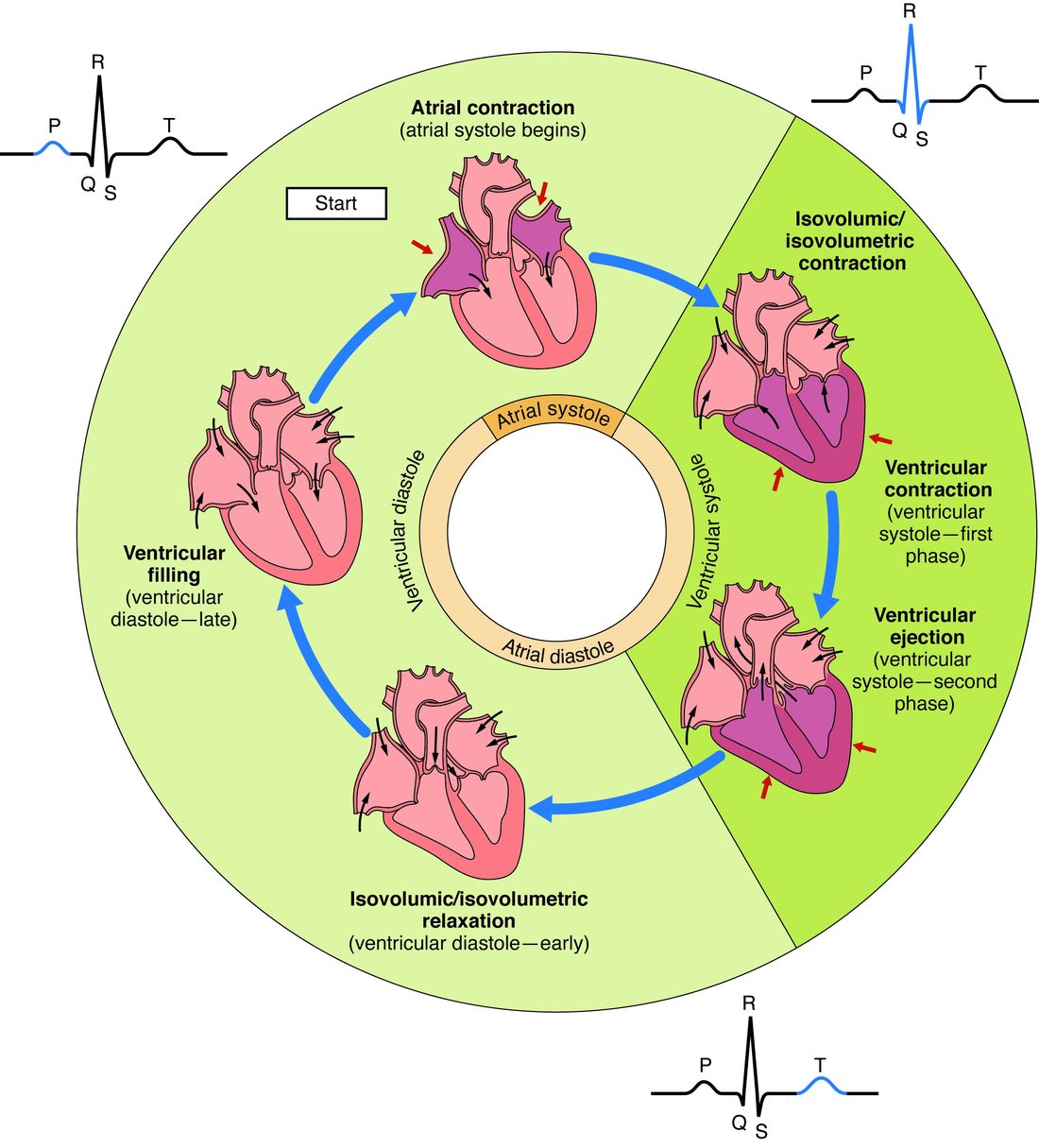
- A wave of depolarization spreads through the ventricles (the QRS complex on the ECG), and ventricular muscle contracts.Ventricular pressure rises above atrial pressure, so the mitral valve closes: S1, and the start of isovolumetric contraction.
- With both valves closed, the contracting ventricle squeezes a fixed volume of blood.Pressure climbs steeply until it passes aortic pressure, about 80 mm Hg, and the aortic valve opens.
- Blood flows down the pressure gradient from ventricle to aorta.Volume falls from EDV (about 120 mL) to ESV (about 50 mL): a stroke volume of about 70 mL.
- The ventricle relaxes and its pressure falls below aortic pressure.Blood slips back, fills the aortic cusps and closes the valve: S2, the dicrotic notch, and the start of isovolumetric relaxation.
- Ventricular pressure keeps falling until it drops below atrial pressure.The mitral valve opens and the ventricle fills, mostly passively, until the atria contract to top it off.
6Core concepts
7A common mistake
The wrong idea: The heart sounds are made by the valve cusps slapping together, or by the heart muscle contracting.
What actually happens: Muscle contraction is almost silent through a stethoscope, and the thin cusps are too soft to clap. Each sound comes from moving blood being stopped abruptly as a set of valves closes, which sets the blood, cusps and chamber walls vibrating together. That is why S1 is timed exactly to atrioventricular valve closure and S2 to semilunar valve closure.
8Check yourself
Anything you miss goes into your review queue.
1. What makes the aortic valve open?
- The papillary muscles pull its cusps apart
- Left ventricular pressure rises above aortic pressure
- A nerve signal arriving with the QRS complex
- Aortic pressure rises above left ventricular pressure
Show the answer
Valves open and close only because of the pressure difference across them. When pressure behind the aortic valve (in the ventricle) exceeds pressure in front of it (in the aorta), blood pushes the cusps aside.
- The papillary muscles pull its cusps apart: Papillary muscles attach to the atrioventricular valves, not the semilunar valves, and they never pull any valve open.
- Correct: Left ventricular pressure rises above aortic pressure: Correct. The valve opens when ventricular pressure exceeds aortic pressure, at about 80 mm Hg at rest.
- A nerve signal arriving with the QRS complex: Valves have no muscle and receive no nerve signals. The QRS complex triggers ventricular contraction, which raises pressure; pressure then opens the valve.
- Aortic pressure rises above left ventricular pressure: Higher pressure in the aorta than in the ventricle pushes blood backward into the cusps, which closes the valve.
2. Use the graph. At the moment the aortic valve opens, about what is the left ventricular pressure?
- About 10 mm Hg
- About 120 mm Hg
- About 80 mm Hg
- About 100 mm Hg
Show the answer
The aortic valve opens where the rising ventricular trace crosses the aortic trace. Aortic pressure has fallen to its lowest value, about 80 mm Hg, so that is where the crossing happens.
- About 10 mm Hg: A few mm Hg, near the bottom of the graph, is where the mitral valve closes, as ventricular pressure first rises above atrial pressure.
- About 120 mm Hg: 120 mm Hg is the peak reached during ejection, after the valve is already open.
- Correct: About 80 mm Hg: Correct. Ventricular pressure must exceed aortic pressure, about 80 mm Hg, before the valve opens.
- About 100 mm Hg: About 100 mm Hg is near where the aortic valve closes at the end of ejection, not where it opens.
3. Starting with atrial contraction, put the phases of the cardiac cycle in order.
- Atrial contraction
- Isovolumetric contraction
- Ventricular ejection
- Isovolumetric relaxation
- Ventricular filling
Show the answer
The atria top off the ventricles; the ventricles contract with both valves closed until the aortic valve opens; blood is ejected; the aortic valve closes and pressure falls with both valves closed; the mitral valve opens and the ventricles fill until the atria contract again.
- Correct order: 1. Atrial contraction 2. Isovolumetric contraction 3. Ventricular ejection 4. Isovolumetric relaxation 5. Ventricular filling
4. A left ventricle holds 150 mL at the end of diastole and ejects 60 mL per beat. What is its ejection fraction?
- 40 percent
- 60 percent
- 90 percent
- 25 percent
Show the answer
EF = SV ÷ EDV × 100 = 60 ÷ 150 × 100 = 40 percent. That is clearly below the normal range of about 50 to 70 percent.
- Correct: 40 percent: Correct. 60 ÷ 150 = 0.40.
- 60 percent: This is the fraction left behind: ESV = 150 − 60 = 90 mL, and 90 ÷ 150 = 60 percent.
- 90 percent: This is the ESV in mL (150 − 60 = 90), not a percentage ejected.
- 25 percent: This divides 60 by 240, which has no meaning here.
5. Which statements describe the left ventricle during isovolumetric relaxation? Select all that apply.
- Both the mitral and aortic valves are closed
- Ventricular pressure is falling steeply
- Ventricular volume stays at the end-systolic volume
- Blood is flowing from the ventricle into the aorta
- The mitral valve is open
Show the answer
The aortic valve has just closed (S2) and the mitral valve has not yet opened, so the relaxing ventricle loses pressure with no change in volume, which stays at ESV.
- Correct: Both the mitral and aortic valves are closed: True. Aortic pressure is above ventricular pressure, and ventricular pressure is still above atrial pressure.
- Correct: Ventricular pressure is falling steeply: True. The muscle is relaxing inside a sealed chamber.
- Correct: Ventricular volume stays at the end-systolic volume: True. No blood can enter or leave until the mitral valve opens.
- Blood is flowing from the ventricle into the aorta: False. The aortic valve closed at the start of this phase, so ejection has ended.
- The mitral valve is open: False. The mitral valve opens only when ventricular pressure falls below atrial pressure, which ends this phase.
6. Mr. Abara, 81, gets short of breath climbing stairs. His doctor hears a harsh whooshing sound that starts after S1 and ends before S2, loudest just right of the upper sternum. An ultrasound shows stiff, calcified aortic valve cusps. What best explains the sound?
- Blood leaking back into the left ventricle while it relaxes
- Blood forced at high speed through a narrowed aortic valve during ejection
- The mitral valve failing to close at the start of systole
- Blood rushing into the left ventricle through the mitral valve as it fills early in diastole
Show the answer
The murmur falls between S1 and S2, so it happens during ventricular systole. A narrowed (stenotic) aortic valve makes blood jet through a small opening during ejection, and the turbulence is heard as a murmur, best at the aortic listening spot.
- Blood leaking back into the left ventricle while it relaxes: Backflow through a leaky aortic valve happens in diastole, after S2, not between S1 and S2.
- Correct: Blood forced at high speed through a narrowed aortic valve during ejection: Correct. This is aortic stenosis.
- The mitral valve failing to close at the start of systole: A leaky mitral valve also gives a murmur during systole, but it is heard best at the apex, and his ultrasound shows the aortic valve is the problem.
- Blood rushing into the left ventricle through the mitral valve as it fills early in diastole: Early filling happens in diastole, after S2. Normal filling is usually silent.
7. On a left ventricular pressure–volume loop, one side is a vertical line that rises from about 10 to 80 mm Hg at the loop's largest volume. Which phase does it show?
- Ventricular filling
- Ventricular ejection
- Isovolumetric contraction
- Isovolumetric relaxation
Show the answer
A vertical line means pressure changes with no change in volume. At the largest volume (EDV), with pressure rising, that is isovolumetric contraction, between mitral valve closure and aortic valve opening.
- Ventricular filling: During filling, volume rises, so the line runs across the bottom of the loop, not up the side.
- Ventricular ejection: During ejection volume falls, so that side runs across the top from right to left.
- Correct: Isovolumetric contraction: Correct. Rising pressure at constant maximum volume is isovolumetric contraction.
- Isovolumetric relaxation: Isovolumetric relaxation is also vertical, but pressure falls and it sits at the smallest volume (ESV), on the left side.
9Summary
Valves open and close only because of the pressure difference across them. In each cycle the ventricles fill mostly passively, are topped off by atrial contraction, contract with both valves closed (isovolumetric contraction) until the aortic valve opens, eject blood, and relax with both valves closed (isovolumetric relaxation) until the mitral valve opens. S1 (lub) is atrioventricular valve closure and S2 (dub) is semilunar valve closure; murmurs are turbulent flow through a narrowed (stenotic) or leaking (regurgitant) valve. Stroke volume = EDV − ESV, about 120 − 50 = 70 mL at rest, and ejection fraction = SV ÷ EDV, about 58 percent. A pressure–volume loop traces one beat counterclockwise; its width is the stroke volume.