Enzyme Structure
Enzymes are biological catalysts, almost all of them proteins.
Part 1 · Hook
Why this matters
A glass of milk holds a sugar called lactose. Most adults on Earth digest it poorly, yet they digest the sugar in table sugar and bread without trouble. The difference is one protein in the small intestine, lactase. It breaks lactose apart and ignores sucrose and maltose, even though all three are two-sugar molecules of almost the same size. How can a protein tell molecules apart that well? The answer is its shape.
Part 2 · Before you start
What this builds on
Part 3 · Prerequisite check
Quick check before you start
1. What mainly holds a folded protein in its three-dimensional (tertiary) shape?
- Interactions between the side chains of its amino acids, such as hydrogen bonds, ionic attractions and nonpolar clustering
- Peptide bonds between the side chains of amino acids that sit next to each other in the chain
- Phosphate groups that link one amino acid to the next along the backbone of the chain
Show the answer
Peptide bonds join amino acids into a chain (primary structure). The 3-D folding comes from side chains attracting or repelling each other: polar with polar, charged with opposite charges, nonpolar ones clustering away from water.
- Correct: Interactions between the side chains of its amino acids, such as hydrogen bonds, ionic attractions and nonpolar clustering:
- Peptide bonds between the side chains of amino acids that sit next to each other in the chain:
- Phosphate groups that link one amino acid to the next along the backbone of the chain:
2. A protein is heated to 80 °C. It unfolds, but its chain of amino acids is still connected in the same order. What has happened?
- It has been hydrolyzed into separate amino acids
- It has been denatured: its shape changed but its sequence did not
- It has gained new amino acids from the solution around it
Show the answer
Denaturation breaks the weak interactions that hold the shape; the peptide bonds of the chain stay intact. Hydrolysis would break the chain itself.
- It has been hydrolyzed into separate amino acids:
- Correct: It has been denatured: its shape changed but its sequence did not:
- It has gained new amino acids from the solution around it:
3. Enzymes speed up hydrolysis. In a hydrolysis reaction, what happens?
- Two monomers are joined and a water molecule is released
- A polymer is built by adding monomers one at a time without water
- A bond between monomers is broken by adding a water molecule
Show the answer
Hydro (water) + lysis (splitting): water is added across a bond, splitting a polymer or a dimer into smaller pieces. Joining monomers and releasing water is dehydration synthesis.
- Two monomers are joined and a water molecule is released:
- A polymer is built by adding monomers one at a time without water:
- Correct: A bond between monomers is broken by adding a water molecule:
Part 4 · See it
See it first
Part 5 · Step by step
How it works, step by step
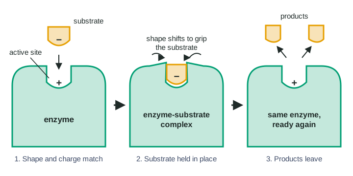
- An enzyme's chain of amino acids folds as its side chains attract and repel each other.The folded protein has a pocket or groove on its surface, the active site, lined by a particular set of side chains.
- The side chains lining the active site give it a specific shape and a specific pattern of charged, polar and nonpolar spots.A molecule binds well only if its shape and charges are complementary to that pattern. That molecule is the enzyme's substrate, and the match is called enzyme specificity.
- The substrate settles into the active site, held by weak attractions such as hydrogen bonds and ionic attractions.An enzyme-substrate complex forms, and the active site shifts slightly to grip the substrate more tightly: induced fit.
- The tight grip holds particular bonds of the substrate close to particular side chains of the enzyme.Those bonds break or form, the substrate becomes product, and the product no longer fits as well, so it is released.
- The enzyme comes out of the reaction with the same shape it started with.Its active site is free to bind another substrate molecule, over and over: the enzyme is a catalyst.
- Anything that changes the folding, such as high heat, an extreme pH or a swapped amino acid in the active site, changes the active site's shape or charges.The substrate no longer fits or is no longer attracted, so little or no product is made.
Part 6 · Key ideas
Key ideas
- An enzyme is a catalyst: it speeds up a reaction and comes out unchanged. Almost all enzymes are proteins; a few are RNA molecules.
- The active site is a small part of a large protein, but the whole protein's folding creates it. A change far from the active site can still matter if it changes how the protein folds.
- Specificity needs two matches: shape (the substrate fits) and chemistry (charges and polarity attract rather than repel). Data that change one but not the other let you tell them apart.
- Induced fit: the active site is flexible. Binding the right substrate changes the enzyme's shape slightly; the old lock-and-key picture treated it as rigid.
Part 7 · Misconception
A common mistake
The wrong idea: An enzyme acts on its substrate because the substrate is the right size, so any molecule of the same size will be acted on.
What actually happens: Size is not enough. The substrate must match the active site's shape in detail and its pattern of charges and polar groups. Lactose and maltose are the same size, but lactase acts only on lactose.
Part 8 · Check yourself
Check yourself
Exam-style questions. Anything you miss goes into your review queue.
Experimental setup
Does enzyme H change shape?
Enzyme H is built from two lobes with the active site in the cleft between them. H joins sugar G to a second molecule. Researchers used a method that measures the distance between two points, one on each lobe, while different molecules were added. Water is present in every sample and is small enough to enter the cleft.
| Molecule added | Description | Gap between lobes (nm) | Product made? |
|---|---|---|---|
| None (water only) | — | 2.6 | no |
| Sugar G | the substrate | 0.9 | yes |
| Molecule L | same size as G; one –OH group of G replaced by –H | 2.3 | no |
| Molecule M | about twice the size of G | 2.6 | no |
1. Which model of substrate binding do the data best support?
- Induced fit: the lobes close around the substrate when it binds, so the enzyme's shape changes during binding.
- Lock and key: the active site has a fixed shape, and the substrate fits it without the enzyme moving.
- The substrate changes shape to fit the enzyme, while the enzyme's lobes stay where they were.
- The enzyme changes shape in the same way whenever any molecule that is small enough to enter the cleft is present.
Show the answer
The gap shrinks from 2.6 nm to 0.9 nm only when G binds. The enzyme itself changes shape as the substrate binds: induced fit.
- Correct: Induced fit: the lobes close around the substrate when it binds, so the enzyme's shape changes during binding.: The lobes move closer by 1.7 nm when G binds, the enzyme shape change that defines induced fit.
- Lock and key: the active site has a fixed shape, and the substrate fits it without the enzyme moving.: A rigid lock would keep the gap the same with or without G; the gap clearly changes.
- The substrate changes shape to fit the enzyme, while the enzyme's lobes stay where they were.: The measurement is between two points on the enzyme, so a change in that distance shows the enzyme moving.
- The enzyme changes shape in the same way whenever any molecule that is small enough to enter the cleft is present.: Water is in every sample and is small enough to enter, yet the gap stays at 2.6 nm; molecule L also leaves it mostly open.
2. Molecule L is the same size as G but lacks one –OH group. The gap barely closes and no product forms. Which explanation is most consistent with these data?
- Without that –OH, a hydrogen bond to an active-site side chain is lost, so binding does not trigger the full shape change.
- Molecule L is too large to enter the cleft, so it stays outside and the lobes remain open.
- Molecule L binds the active site more tightly than sugar G does, and this tight binding locks the two lobes in their open position.
- Molecule L has a positive charge that repels the lobes and pushes them farther apart.
Show the answer
An –OH group is polar and can hydrogen bond with a polar side chain. Replacing it with –H removes that contact. With less of the right attraction, the lobes do not close and the substrate's bonds are not held in place to react.
- Correct: Without that –OH, a hydrogen bond to an active-site side chain is lost, so binding does not trigger the full shape change.: Losing one polar contact is enough to stop the shape change, showing that both shape and chemical matching matter.
- Molecule L is too large to enter the cleft, so it stays outside and the lobes remain open.: The table says L is the same size as G, so it can enter the cleft as G does.
- Molecule L binds the active site more tightly than sugar G does, and this tight binding locks the two lobes in their open position.: Nothing in the data shows tighter binding; an open gap fits weak, partial binding better.
- Molecule L has a positive charge that repels the lobes and pushes them farther apart.: Swapping –OH for –H adds no charge, and the gap (2.3 nm) is smaller than with water alone, not larger.
3. A variant of enzyme H has its lobes stuck at a 0.9 nm gap before any substrate is added. What is the most likely effect on product formation, and why?
- It falls, because sugar G can no longer get into the closed cleft to reach the active site.
- It rises, because the enzyme is already in the shape it takes when the substrate is bound.
- It stays the same, because the gap matters after the product has been released, not during binding.
- It rises, because water molecules can no longer enter the cleft and compete with G for space there.
Show the answer
Closing happens around the substrate. If the cleft is already closed, G cannot enter, so the enzyme-substrate complex cannot form and little product is made.
- Correct: It falls, because sugar G can no longer get into the closed cleft to reach the active site.: The substrate must get in before the lobes close; a cleft that is already shut keeps it out.
- It rises, because the enzyme is already in the shape it takes when the substrate is bound.: Being in the bound shape is useless without the substrate inside; the closed lobes block entry.
- It stays the same, because the gap matters after the product has been released, not during binding.: In the data the gap changes when G binds, before product forms, so it matters for binding.
- It rises, because water molecules can no longer enter the cleft and compete with G for space there.: Water entering the open cleft did not stop the original enzyme from working, so keeping it out offers no gain.
4. A student claims the active site of enzyme H is a rigid mold that fits G exactly. Which observation in the table most directly contradicts the claim?
- The gap between the lobes shrinks from 2.6 nm to 0.9 nm when G is added.
- Molecule M, about twice the size of G, makes no product.
- Water is present in each of the samples but is not joined to anything.
- Each gap in the table is the mean of six measurements rather than one.
Show the answer
A rigid mold would not move. The 1.7 nm change shows the enzyme's shape changes when G binds.
- Correct: The gap between the lobes shrinks from 2.6 nm to 0.9 nm when G is added.: This is direct evidence that the enzyme's shape changes on binding, which a rigid mold could not do.
- Molecule M, about twice the size of G, makes no product.: A molecule too big to fit makes no product under either model, so this cannot tell them apart.
- Water is present in each of the samples but is not joined to anything.: Water gives no product under either model; it says nothing about whether the enzyme moves.
- Each gap in the table is the mean of six measurements rather than one.: Averaging repeated measurements makes the data more reliable but says nothing about rigidity.
5. An enzyme's active site is lined by a positively charged side chain at its base and a patch of nonpolar side chains along one wall. Which molecule is most likely to be its substrate?
- A molecule with a negatively charged group at one end and a nonpolar ring along one side
- A molecule with a positively charged group at one end and a nonpolar ring along one side
- A molecule with a negatively charged group at one end and several polar –OH groups along one side
- A molecule with no charged groups and several polar –OH groups spread over its surface
Show the answer
A negative group is attracted to the positive side chain, and a nonpolar ring clusters with the nonpolar wall. Both parts of the substrate match the active site.
- Correct: A molecule with a negatively charged group at one end and a nonpolar ring along one side: Both regions match: opposite charges attract and nonpolar groups cluster together.
- A molecule with a positively charged group at one end and a nonpolar ring along one side: A positive group would be repelled by the positive side chain at the base.
- A molecule with a negatively charged group at one end and several polar –OH groups along one side: The charge matches, but polar –OH groups would sit against a nonpolar wall instead of a matching surface.
- A molecule with no charged groups and several polar –OH groups spread over its surface: With no charged group, nothing in this molecule is attracted to the positive side chain, and its polar groups do not match the nonpolar wall.
6. A human enzyme is kept at 70 °C for 10 minutes and then cooled to 37 °C. Its substrate is added, and almost no product forms. Which explanation is best?
- Heat broke weak bonds holding the protein's fold, and the active site did not regain its shape on cooling.
- The heat broke the peptide bonds of the enzyme, leaving free amino acids that are unable to bind the substrate.
- At 37 °C the molecules move too slowly for the substrate to reach an enzyme that was heated.
- The heat used up the enzyme's stored energy, so it has no energy left to change the substrate into product.
Show the answer
High heat denatures the protein: it breaks hydrogen bonds and other weak interactions that hold the fold. Here the shape did not come back on cooling, so the active site no longer fits the substrate.
- Correct: Heat broke weak bonds holding the protein's fold, and the active site did not regain its shape on cooling.: Denaturation unfolds the protein and wrecks the active site's shape; this enzyme did not refold.
- The heat broke the peptide bonds of the enzyme, leaving free amino acids that are unable to bind the substrate.: Heating to 70 °C unfolds proteins but does not hydrolyze peptide bonds; the chain stays intact.
- At 37 °C the molecules move too slowly for the substrate to reach an enzyme that was heated.: 37 °C is the normal temperature for a human enzyme, so slow movement cannot be the reason.
- The heat used up the enzyme's stored energy, so it has no energy left to change the substrate into product.: Enzymes do not store energy that runs out; they work by holding the substrate in the active site.
7. A drug molecule has nearly the same shape and charge pattern as an enzyme's substrate, so it binds the active site, but the enzyme cannot change it into product. The drug is added to a tube of enzyme and substrate. Predict the change in each variable.
| Variable | Change |
|---|---|
| Number of active sites holding the substrate at any moment | — |
| Product formed per minute | — |
| Total number of enzyme molecules in the tube | — |
Show the answer
A look-alike molecule takes up active sites without reacting, so fewer sites are free for the real substrate and product forms more slowly. The enzymes themselves are still there.
- Number of active sites holding the substrate at any moment: decreases. Some active sites are occupied by the drug, so fewer are free to hold the substrate at a given moment.
- Product formed per minute: decreases. Product forms only from substrate in an active site; with fewer sites holding substrate, less product forms each minute.
- Total number of enzyme molecules in the tube: no change. The drug binds the enzymes but does not destroy them or add new ones, so the count is unchanged.
Part 9 · Summary
Summary
Enzymes are biological catalysts, almost all of them proteins. Folding gives each enzyme an active site whose shape and pattern of charged, polar and nonpolar side chains match one substrate, or a few similar ones: that is specificity. The substrate binds by weak attractions to form the enzyme-substrate complex, and the active site shifts to grip it (induced fit). The products are released and the unchanged enzyme can act again. Anything that changes the active site's shape or charges, from heat to a single swapped amino acid, changes what the enzyme can bind.
Part 10 · Up next
What comes next
Part 11 · Connections