T cells and cell-mediated immunity
1Why this matters
Mr. Okafor, 52, received a kidney from a deceased donor eight weeks ago. He ran out of his tacrolimus tablets on a trip and skipped them for five days. Now he has a low fever, the new kidney is swollen and tender, and he is making much less urine. A biopsy shows the kidney packed with his own T cells, killing the donor cells one by one. To see why one missed drug lets this happen, you need to know how T cells are switched on and what each kind does.
2What this builds on
3Quick check before you start
1. Which cells carry MHC class I proteins?
- Only antigen-presenting cells
- Nearly every cell with a nucleus
- Only T cells and B cells
Show the answer
MHC class I proteins sit on nearly every nucleated cell and display peptides made inside that cell. MHC class II proteins are the ones found mainly on antigen-presenting cells.
- Only antigen-presenting cells:
- Correct: Nearly every cell with a nucleus:
- Only T cells and B cells:
2. What is apoptosis?
- Programmed cell death, in which a cell dismantles itself into tidy fragments
- Bursting of a cell after water rushes in
- Division of a cell into two identical daughters
Show the answer
Apoptosis is programmed cell death: enzymes inside the cell take it apart into membrane-wrapped fragments that phagocytes engulf, with little inflammation.
- Correct: Programmed cell death, in which a cell dismantles itself into tidy fragments:
- Bursting of a cell after water rushes in:
- Division of a cell into two identical daughters:
3. A lymphocyte whose receptor fits one antigen meets that antigen and multiplies into many identical cells. What is this called?
- Negative selection
- Clonal selection and expansion
- Antigen processing
Show the answer
Clonal selection picks out the rare lymphocyte whose receptor fits the antigen; clonal expansion is its division into a clone of identical cells.
- Negative selection:
- Correct: Clonal selection and expansion:
- Antigen processing:
4Anatomy
With labels hidden, select a box to reveal its label.
5How it works, step by step
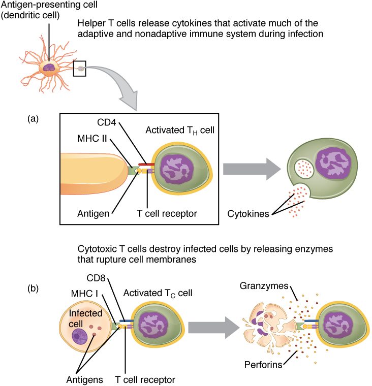
- A dendritic cell engulfs a virus at the site of infection, senses it, and carries viral peptides in its MHC proteins to a lymph node.It displays those peptides and makes B7 proteins on its surface.
- A naïve T cell whose receptor fits one of the peptides binds it (signal 1), and its CD28 binds the B7 (costimulation, signal 2).The T cell is activated and starts making IL-2 and IL-2 receptor proteins.
- IL-2 acts on the same cell and its neighbors.The T cell divides again and again: clonal expansion into tens of thousands to millions of identical effector T cells and some memory T cells.
- Effector cytotoxic T cells leave the node and find infected cells showing the viral peptide in MHC class I.They release perforin and granzymes and engage Fas, switching on the target's caspases.
- Caspases take the infected cell apart from inside.The cell dies by apoptosis before it can release new virus, and macrophages engulf the fragments.
6Core concepts
7A common mistake
The wrong idea: T cells recognize a virus or bacterium directly, the way antibodies do.
What actually happens: A T cell receptor cannot bind a free microbe or a whole protein. It binds only a short peptide held in an MHC protein on another cell's surface. That is why T cells need antigen-presenting cells to be activated, why cytotoxic T cells find infected cells rather than free virus, and why a cell that stops making MHC class I becomes invisible to them.
8Check yourself
Anything you miss goes into your review queue.
1. A liver cell infected with a virus makes viral proteins and displays peptides from them on its surface. Which T cell recognizes it, and through which MHC protein?
- A CD4 T cell, through MHC class II
- A CD8 T cell, through MHC class I
- A CD4 T cell, through MHC class I
- A CD8 T cell, through MHC class II
Show the answer
Peptides made inside a cell are displayed on MHC class I, which nearly every nucleated cell carries. CD8 binds MHC class I, so a CD8 cytotoxic T cell recognizes the infected cell and kills it.
- A CD4 T cell, through MHC class II: A liver cell is not an antigen-presenting cell and carries little MHC class II. Class II shows engulfed material, not proteins the cell makes itself.
- Correct: A CD8 T cell, through MHC class I: Correct. Internal (here viral) peptides go on MHC class I, and CD8 pairs with class I.
- A CD4 T cell, through MHC class I: CD4 binds MHC class II, not class I, so a CD4 T cell cannot lock onto this complex.
- A CD8 T cell, through MHC class II: The pairing is reversed. CD8 binds class I; MHC class II is recognized with CD4.
2. Mr. Okafor has a transplanted kidney and takes tacrolimus, which stops activated T cells from making IL-2. He skips it for five days, and acute rejection begins. What did tacrolimus normally prevent?
- His B cells from making antibodies against ABO antigens
- His T cells from recognizing donor HLA proteins
- His neutrophils from entering the kidney
- His T cells from expanding into large clones
Show the answer
Activated T cells make IL-2, which drives them to divide again and again. Without IL-2, T cells that recognize donor HLA cannot build the large clones of effector cells that destroy the graft. Off the drug, clonal expansion resumes and rejection follows.
- His B cells from making antibodies against ABO antigens: Acute rejection is driven by T cells, and ABO antibodies cause hyperacute rejection within minutes. The drug acts on T cell IL-2.
- His T cells from recognizing donor HLA proteins: Recognition (signal 1) still happens on the drug. What is blocked is the IL-2 that turns recognition into expansion.
- His neutrophils from entering the kidney: Neutrophils are innate cells and do not use IL-2 to expand. The drug's target is T cell activation.
- Correct: His T cells from expanding into large clones: Correct. Blocking IL-2 blocks clonal expansion of the T cells that react to donor HLA.
3. Put the steps in order, from a microbe entering the skin to effector T cells leaving the lymph node.
- A dendritic cell engulfs the microbe in the skin
- The dendritic cell travels to a lymph node and displays microbial peptides in MHC
- A naïve T cell with a matching T cell receptor binds a peptide–MHC complex
- CD28 on the T cell binds B7 on the dendritic cell
- The T cell makes IL-2 and divides into a clone
- Effector T cells leave the node for the infected tissue
Show the answer
The dendritic cell captures antigen at the site of infection, carries it to the lymph node and presents it. A matching naïve T cell receives signal 1 (recognition) and signal 2 (costimulation), makes IL-2, expands, and its effector daughters leave for the tissue.
- Correct order: 1. A dendritic cell engulfs the microbe in the skin 2. The dendritic cell travels to a lymph node and displays microbial peptides in MHC 3. A naïve T cell with a matching T cell receptor binds a peptide–MHC complex 4. CD28 on the T cell binds B7 on the dendritic cell 5. The T cell makes IL-2 and divides into a clone 6. Effector T cells leave the node for the infected tissue
4. Select every mechanism a cytotoxic T cell uses to kill a virus-infected cell.
- Releasing perforin, which makes pores in the target's membrane
- Releasing antibodies that coat the target
- Delivering granzymes that switch on the target's caspases
- Engulfing the target whole
- Binding Fas on the target with Fas ligand
- Releasing histamine onto the target
Show the answer
Cytotoxic T cells kill by contact. Perforin opens pores, granzymes enter and activate caspases, and Fas ligand triggers the same caspases through Fas. Each route ends in apoptosis.
- Correct: Releasing perforin, which makes pores in the target's membrane: Correct. Perforin forms the pores through which granzymes enter.
- Releasing antibodies that coat the target: T cells do not make antibodies; those come from cells descended from B cells.
- Correct: Delivering granzymes that switch on the target's caspases: Correct. Granzymes activate caspases, which carry out apoptosis.
- Engulfing the target whole: Engulfing is what phagocytes such as macrophages do. The cytotoxic T cell makes its target die by apoptosis, and a macrophage then engulfs the fragments.
- Correct: Binding Fas on the target with Fas ligand: Correct. Fas ligand binding Fas is the granule-free route to apoptosis.
- Releasing histamine onto the target: Histamine is released by mast cells and basophils in inflammation, not by T cells.
5. A student described how a cytotoxic T cell kills an infected cell. One step is wrong. Which one?
- The T cell receptor and CD8 bind viral peptide in MHC class I
- The T cell turns its granules toward the contact point
- Perforin forms pores in the target's membrane
- Granzymes enter and switch on the target's caspases
- The target cell swells and bursts, spilling its contents into the tissue
Show the answer
Granzymes and caspases make the target die by apoptosis. It breaks up into tidy fragments that macrophages engulf, so its contents, including virus being assembled inside, are not spilled and there is little inflammation.
- The T cell receptor and CD8 bind viral peptide in MHC class I: This step is right. Recognition needs the peptide in MHC class I, bound by the T cell receptor and CD8.
- The T cell turns its granules toward the contact point: This step is right. Aiming the granules keeps the damage to the one cell being attacked.
- Perforin forms pores in the target's membrane: This step is right. Perforin opens the way for granzymes.
- Granzymes enter and switch on the target's caspases: This step is right. Granzymes trigger the target's own caspases.
- Correct: The target cell swells and bursts, spilling its contents into the tissue: This is the error. The target dies by apoptosis, not by bursting.
6. A child has a heavy roundworm infection of the gut. Her blood shows many eosinophils. Which helper T cell subset is driving this response?
- Th1 cells releasing interferon gamma
- Regulatory T cells releasing IL-10
- Cytotoxic T cells releasing granzymes onto the worm
- Th2 cells releasing IL-4, IL-5 and IL-13
Show the answer
Th2 cells direct the response to parasitic worms. IL-5 calls up eosinophils, IL-13 increases mucus and speeds turnover of the gut lining, and IL-4 steers B cells and promotes further Th2 development.
- Th1 cells releasing interferon gamma: Th1 cells arm macrophages and cytotoxic T cells against microbes that live inside cells. Interferon gamma actually holds Th2 development back.
- Regulatory T cells releasing IL-10: Regulatory T cells damp responses down; they do not raise eosinophil numbers.
- Cytotoxic T cells releasing granzymes onto the worm: Cytotoxic T cells are not helper T cells, and a worm is far too large to be killed by granzymes.
- Correct: Th2 cells releasing IL-4, IL-5 and IL-13: Correct. Worms trigger a Th2 response, and IL-5 raises eosinophils.
7. A kidney transplanted into a patient turns dark and stops working within minutes of the clamps being released, as clots form in its vessels. What caused this?
- Preexisting antibodies against the donor's vessels
- Recipient cytotoxic T cells killing the kidney's cells one by one
- Donor T cells attacking the patient
- Slow scarring of the graft's arteries
Show the answer
Rejection within minutes is hyperacute rejection. Only antibodies already present, for example against the donor's ABO antigens or HLA, can act that fast. They bind the graft's blood vessel lining, triggering clotting. ABO matching and crossmatching are done to prevent it.
- Correct: Preexisting antibodies against the donor's vessels: Correct. Preformed antibodies cause hyperacute rejection.
- Recipient cytotoxic T cells killing the kidney's cells one by one: T cell rejection needs activation and clonal expansion first, which takes days, not minutes.
- Donor T cells attacking the patient: Donor T cells attacking the recipient is graft-versus-host disease, which follows marrow transplants and takes weeks.
- Slow scarring of the graft's arteries: Scarring of graft arteries is chronic rejection, over months to years.
9Summary
T cells see only peptides held in MHC proteins. CD4 T cells (helper and regulatory) answer MHC class II; CD8 T cells (cytotoxic) answer MHC class I. A naïve T cell is activated in a lymph node by a dendritic cell that provides recognition (signal 1), costimulation through B7 and CD28 (signal 2) and cytokines (signal 3); recognition without costimulation causes anergy. IL-2 drives clonal expansion into effector and memory T cells. Helper T cells direct B cells, cytotoxic T cells and macrophages with cytokines; Th1 cells fight microbes inside cells and Th2 cells fight worms. Cytotoxic T cells kill infected and abnormal cells by apoptosis with perforin, granzymes and Fas ligand. Regulatory T cells suppress other lymphocytes. Transplant rejection is mainly T cells reacting to foreign HLA, reduced by tissue typing, crossmatching and drugs that block T cell activation; in graft-versus-host disease, donor T cells attack the recipient.