The pectoral girdle and upper limb
1Why this matters
Diego, 20, slips on ice and lands on his outstretched hand. His wrist aches, and pressing the small hollow at the base of his thumb makes him wince. The X-ray looks normal, but the doctor splints his wrist anyway and orders another image in two weeks. The reason is a small boat-shaped wrist bone with an unusual blood supply.
2What this builds on
3Quick check before you start
1. In anatomical position, which way do the palms face?
- Forward
- Toward the body
- Backward
Show the answer
In anatomical position you stand upright with your arms at your sides and your palms facing forward. That is the position that medial and lateral in the forearm and hand refer to.
- Correct: Forward:
- Toward the body:
- Backward:
2. What is a small, rounded bump on a bone called?
- A fossa
- A tubercle
- A foramen
Show the answer
A tubercle is a small rounded bump, usually where a tendon or ligament anchors. A fossa is a shallow hollow, and a foramen is a hole.
- A fossa:
- Correct: A tubercle:
- A foramen:
3. Which division of the skeleton do the arm bones and shoulder bones belong to?
- Axial
- Appendicular
- Neither
Show the answer
The appendicular skeleton is the limbs plus the bones that attach them to the trunk, so it includes the arm bones and the shoulder bones.
- Axial:
- Correct: Appendicular:
- Neither:
4Anatomy
With labels hidden, select a box to reveal its label.
5How it works, step by step
- A person falls onto an outstretched hand with the wrist bent back.The force travels from the palm through the carpals into the radius.
- The scaphoid sits against the radius and bridges the two rows of carpals.It takes much of the load and can snap across its narrow middle.
- Blood vessels enter the scaphoid mainly at its far end.A break across its middle can cut the near piece off from its blood supply.
- Without blood, the bone cells of the near piece die.The break may fail to heal, so a suspected scaphoid fracture is splinted even when the first X-ray looks normal.
6Core concepts
7A common mistake
The wrong idea: Hitting your "funny bone" hurts because you bruised a bone.
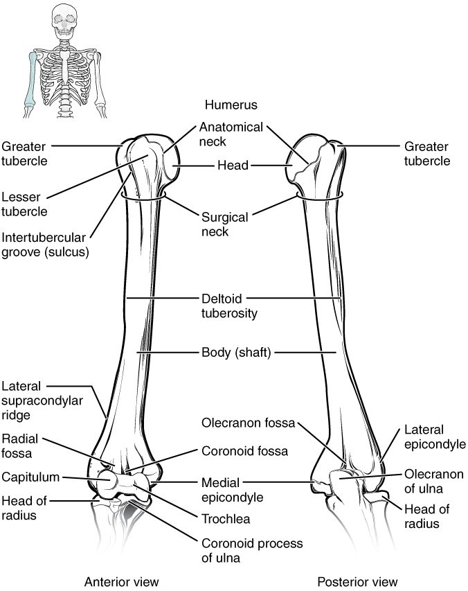
What actually happens: The funny bone is not a bone. A nerve runs in a groove on the back of the medial epicondyle of the humerus, at the inner back of your elbow, with only skin over it. A knock squeezes the nerve against the bone, and the tingling runs along the nerve's path to your little finger.
8Check yourself
Anything you miss goes into your review queue.
1. Name the pinned part of the scapula.
- Coracoid process
- Acromion
- Glenoid cavity
- Spine of the scapula
Show the answer
The acromion is the flat end of the scapula's spine. It forms the bony tip of the shoulder and meets the clavicle.
- Coracoid process: The coracoid process is the hook that points forward under the clavicle.
- Correct: Acromion: Correct. The flat projection at the tip of the shoulder is the acromion.
- Glenoid cavity: The glenoid cavity is the shallow socket on the outer corner, where the humerus fits.
- Spine of the scapula: The spine is the ridge across the back of the scapula; the acromion is its end.
2. Name the pinned narrowing of the humerus.
- Anatomical neck
- Intertubercular groove
- Deltoid tuberosity
- Surgical neck
Show the answer
The surgical neck is the narrowing of the shaft just below the tubercles. It is a common fracture site, especially in older adults.
- Anatomical neck: The anatomical neck is the groove right at the edge of the head, above the tubercles.
- Intertubercular groove: The intertubercular groove is the channel between the tubercles, running up and down.
- Deltoid tuberosity: The deltoid tuberosity is farther down, halfway along the lateral shaft.
- Correct: Surgical neck: Correct. The narrowing below the tubercles is the surgical neck.
3. Name the pinned carpal bone on the thumb side of the proximal row.
- Trapezium
- Lunate
- Scaphoid
- Capitate
Show the answer
The scaphoid (scaph- = boat) is the lateral bone of the proximal row, on the thumb side. It meets the radius.
- Trapezium: The trapezium is in the distal row, at the base of the thumb.
- Lunate: The lunate is next to the scaphoid, toward the little finger.
- Correct: Scaphoid: Correct. The boat-shaped bone on the thumb side of the proximal row is the scaphoid.
- Capitate: The capitate is the large central bone of the distal row.
4. Which bone is the only bony link between the upper limb and the axial skeleton?
- Scapula
- Humerus
- Clavicle
- First rib
Show the answer
The clavicle meets the manubrium of the sternum at one end and the acromion of the scapula at the other. The scapula itself is held to the trunk only by muscles.
- Scapula: The scapula does not meet the vertebral column or the ribs; muscles hold it against the back of the rib cage.
- Humerus: The humerus meets the scapula, not the axial skeleton.
- Correct: Clavicle: Correct. The clavicle is the only bony link.
- First rib: The first rib is part of the axial skeleton itself, not a link to the limb.
5. A 20-year-old falls onto his outstretched hand. He is tender in the hollow at the base of his thumb, but the first X-ray looks normal. His wrist is splinted anyway. Why?
- The scaphoid may be broken, and its blood supply can fail
- The pisiform may be broken, and it will slip out of its tendon
- The lunate is the carpal most often broken, and it heals slowly
- The fifth metacarpal may be broken, and it often shows late
Show the answer
The tender hollow at the base of the thumb lies over the scaphoid, the carpal broken most often in this kind of fall. A scaphoid crack may not show on the first X-ray. Blood vessels enter the scaphoid mainly at its far end, so a break can starve the near piece, which may die and fail to heal. Splinting protects it until a later image settles the question.
- Correct: The scaphoid may be broken, and its blood supply can fail: Correct. A missed scaphoid fracture risks losing the blood supply to part of the bone.
- The pisiform may be broken, and it will slip out of its tendon: The pisiform is on the little-finger side of the palm, not the thumb side, and it does not slip out of its tendon.
- The lunate is the carpal most often broken, and it heals slowly: The scaphoid, not the lunate, is the carpal broken most often.
- The fifth metacarpal may be broken, and it often shows late: The fifth metacarpal is at the little-finger side, usually broken by punching, not tender at the base of the thumb.
6. A 45-year-old who types all day has numbness and tingling in her thumb, index and middle fingers that wakes her at night. What forms the roof of the passage where her nerve is being squeezed?
- The flexor retinaculum
- The hamate
- The trochlear notch
- The head of the ulna
Show the answer
This is carpal tunnel syndrome. The carpals form the floor and walls of the carpal tunnel, and the flexor retinaculum stretches across the top as its roof. When the tendons in the tunnel swell, the tight band cannot give, so the nerve is squeezed.
- Correct: The flexor retinaculum: Correct. The flexor retinaculum is the roof of the carpal tunnel.
- The hamate: The hamate is one of the carpals in the wall of the tunnel, not its roof.
- The trochlear notch: The trochlear notch is on the ulna at the elbow.
- The head of the ulna: The head of the ulna is at the wrist but on the little-finger side, outside the tunnel.
7. A student wrote these statements about the forearm in anatomical position. One is wrong. Which one?
- The ulna is on the thumb side of the forearm
- The ulna is larger at the elbow than at the wrist
- The radius is larger at the wrist than at the elbow
- The radius crosses the ulna when the palm turns down
Show the answer
In anatomical position, with the palm forward, the ulna is on the medial, little-finger side, and the radius is on the lateral, thumb side.
- Correct: The ulna is on the thumb side of the forearm: This is the error. The radius is on the thumb side; the ulna is on the little-finger side.
- The ulna is larger at the elbow than at the wrist: This statement is right. The ulna's big end, with the olecranon and trochlear notch, is at the elbow.
- The radius is larger at the wrist than at the elbow: This statement is right. The radius's wide end carries the wrist.
- The radius crosses the ulna when the palm turns down: This statement is right. The radius swings across the front of the ulna when the palm turns down.
9Summary
The pectoral girdle is the clavicle, the only bony link between the arm and the trunk, and the scapula, with its spine, acromion, coracoid process, glenoid cavity and supraspinous, infraspinous and subscapular fossae. The humerus has a head, anatomical and surgical necks, greater and lesser tubercles around the intertubercular groove, a deltoid tuberosity, and at the elbow the epicondyles, capitulum, trochlea and olecranon fossa. The ulna is medial and large at the elbow; the radius is lateral, large at the wrist, and crosses the ulna when the palm turns down. The hand has 8 carpals, 5 metacarpals and 14 phalanges, and the flexor retinaculum roofs the carpal tunnel.