Layers of the skin
1Why this matters
Priya, 34, slices her fingertip on a mandoline and then scrapes her knee on the same day. The scrape oozes a little clear fluid but barely bleeds; the fingertip cut bleeds freely. A week later the scrape has vanished without a mark, while the cut has left a thin line. The difference is how deep each injury went: the skin is built in layers, and each layer behaves differently when you damage it.
2What this builds on
3Quick check before you start
1. What kind of epithelium covers the surface of your skin?
- Simple squamous epithelium
- Keratinized stratified squamous epithelium
- Transitional epithelium
Show the answer
The epidermis is many layers of cells (stratified) whose surface cells are flat (squamous) and filled with keratin (keratinized), which makes it tough and dry.
- Simple squamous epithelium:
- Correct: Keratinized stratified squamous epithelium:
- Transitional epithelium:
2. Which kind of connective tissue has thick collagen bundles running in many directions?
- Dense irregular connective tissue
- Dense regular connective tissue
- Adipose tissue
Show the answer
Dense irregular connective tissue has collagen bundles in many directions, so it resists pulling from any direction. Dense regular tissue, as in tendons, has them in one direction.
- Correct: Dense irregular connective tissue:
- Dense regular connective tissue:
- Adipose tissue:
3. Which cell junction holds neighboring epithelial cells together like rivets?
- Gap junction
- Tight junction
- Desmosome
Show the answer
Desmosomes anchor neighboring cells to each other through their intermediate filaments, so a sheet of cells resists being pulled apart.
- Gap junction:
- Tight junction:
- Correct: Desmosome:
4Anatomy
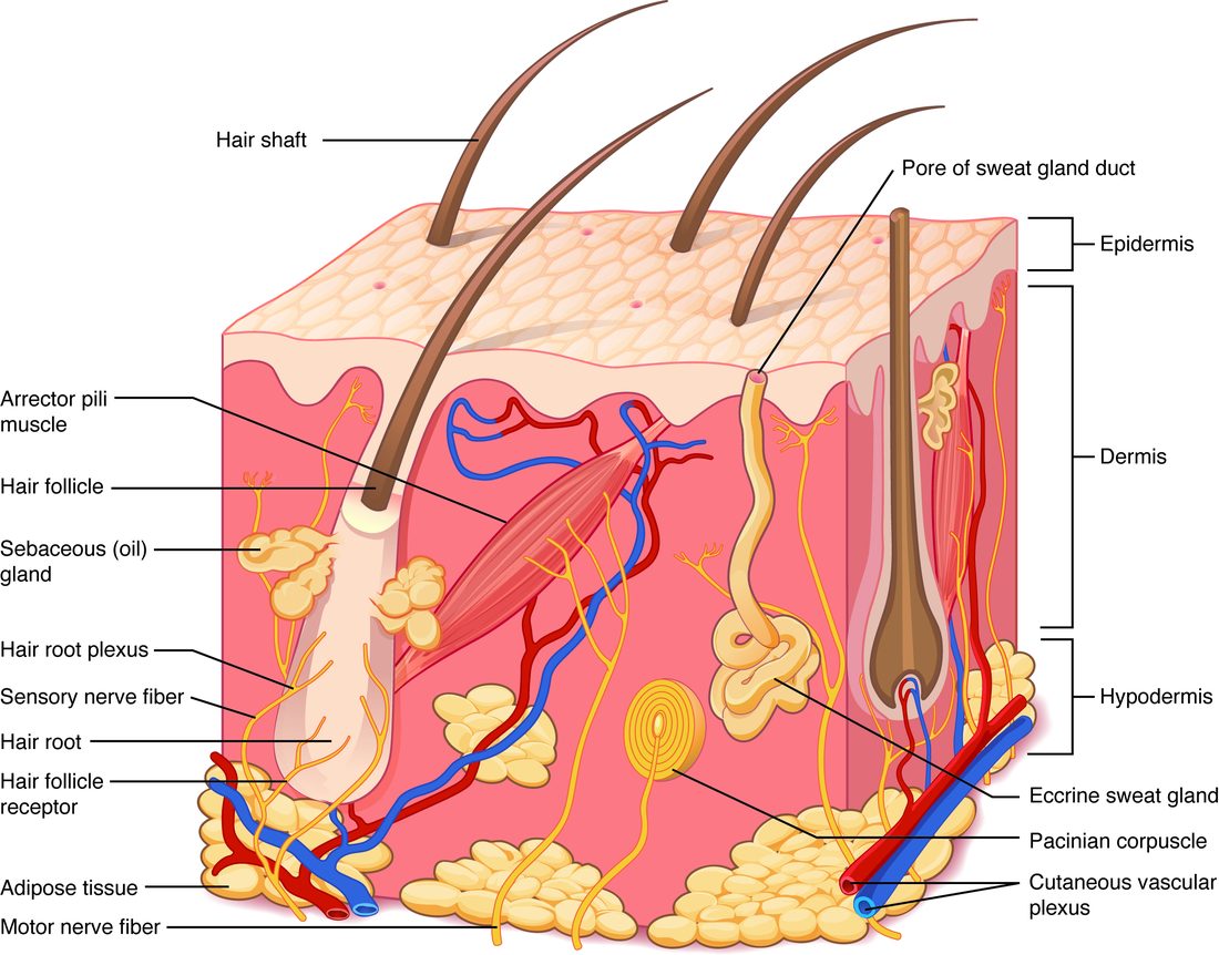
With labels hidden, select a box to reveal its label.
5How it works, step by step
- A basal cell in the stratum basale divides by mitosis.One daughter stays as a stem cell and the other is pushed upward by the cells made beneath it.
- The pushed-up cell enters the stratum spinosum.It becomes a keratinocyte that makes keratin filaments and many desmosomes that bind it to its neighbors.
- The keratinocyte is pushed into the stratum granulosum.Keratohyalin bundles its keratin, lamellar granules release lipid sheets between the cells, and the nucleus and organelles break down, so the cell dies.
- The dead, keratin-filled cell reaches the stratum corneum (passing the stratum lucidum in thick skin).It lies flat among the lipid sheets, part of a tough, water-resistant layer, until the desmosomes holding it are broken down and it is shed.
6Core concepts
7A common mistake
The wrong idea: People with darker skin have more melanocytes.
What actually happens: People of every skin tone have about the same number of melanocytes at a given body site. Skin color comes from what the melanocytes make and hand over: in darker skin the melanosomes are larger, more heavily pigmented, more numerous in the keratinocytes and slower to be broken down. Sun exposure raises melanin output (a tan) without adding melanocytes.
8Check yourself
Anything you miss goes into your review queue.
1. Name the pinned layer.
- Stratum spinosum
- Stratum basale
- Stratum granulosum
- Stratum corneum
Show the answer
The stratum basale is the deepest layer of the epidermis: a single row of cells resting on the basement membrane, where the dermis bulges up into it. Its basal cells divide throughout life, and melanocytes and tactile cells sit among them.
- Stratum spinosum: The stratum spinosum lies just above the pinned layer and is 8 to 10 rows of cells thick, not a single row on the dermis.
- Correct: Stratum basale: Correct. The single deepest row, touching the dermis, is the stratum basale.
- Stratum granulosum: The stratum granulosum lies higher up, where cells flatten and fill with granules.
- Stratum corneum: The stratum corneum is the surface layer of flat dead cells, at the opposite end of the epidermis.
2. A keratinocyte forms in the palm and is pushed toward the surface. Put the layers it passes through in order, starting where it is made.
- Stratum basale
- Stratum spinosum
- Stratum granulosum
- Stratum lucidum
- Stratum corneum
Show the answer
Basal cells divide in the stratum basale. The new keratinocyte makes keratin in the stratum spinosum, forms granules and loses its nucleus in the stratum granulosum, passes through the clear stratum lucidum (present in the thick skin of the palm), and ends as a dead flat cell in the stratum corneum before it is shed.
- Correct order: 1. Stratum basale 2. Stratum spinosum 3. Stratum granulosum 4. Stratum lucidum 5. Stratum corneum
3. Ana gets a paper cut on her finger. It stings, but it does not bleed. Which statement best explains why?
- The cut stayed in the epidermis, which has no blood vessels
- Keratin in the cut cells plugs the torn vessels right away
- The paper edge seals the tiny vessels it cuts by friction heat
- The stratum corneum contains clotting cells
Show the answer
The epidermis is avascular: its cells are fed by diffusion from capillaries in the dermis. A cut that stays in the epidermis reaches no vessel, so it does not bleed. It can still sting, because some nerve endings reach into the lower epidermis.
- Correct: The cut stayed in the epidermis, which has no blood vessels: Correct. No blood vessels in the epidermis means a shallow cut does not bleed.
- Keratin in the cut cells plugs the torn vessels right away: Keratin is a structural protein inside keratinocytes. It plays no part in stopping blood flow.
- The paper edge seals the tiny vessels it cuts by friction heat: A paper cut makes no significant heat. The reason is where the cut stops, not how it seals.
- The stratum corneum contains clotting cells: The stratum corneum is dead keratin-filled cells. Clotting depends on platelets and proteins in the blood, which the epidermis does not contain.
4. Which features belong to thick skin? Select all that apply.
- A stratum lucidum
- Friction ridges
- Hair on its surface
- A very thick stratum corneum
- A thicker dermis than any thin skin
Show the answer
Thick skin, on the palms and soles, has five strata including the stratum lucidum, a very thick stratum corneum, friction ridges from rows of dermal papillae, and no hair. "Thick" describes its epidermis, not its dermis.
- Correct: A stratum lucidum: Yes. Only thick skin has the clear stratum lucidum.
- Correct: Friction ridges: Yes. Rows of dermal papillae push thick skin into friction ridges, the fingerprints of the fingers.
- Hair on its surface: No. Thick skin is hairless.
- Correct: A very thick stratum corneum: Yes. The stratum corneum of thick skin is many times deeper than in thin skin.
- A thicker dermis than any thin skin: No. The skin of the upper back has a deeper dermis than the palm, yet it is thin skin. The name refers to the epidermis.
5. Two adults of similar age, one with very dark skin and one with very light skin, have skin samples taken from the same body site. What is the main difference you would expect to find?
- The dark sample has several times as many melanocytes
- The light sample has melanocytes only in the dermis
- The dark sample has larger, darker melanosomes
- The light sample has no melanin at all
Show the answer
People of all skin tones have about the same number of melanocytes at a given body site. Skin color depends on what those melanocytes make and pass on: in darker skin the melanosomes are larger, more heavily pigmented, more numerous in the keratinocytes and slower to be broken down.
- The dark sample has several times as many melanocytes: Melanocyte numbers are similar across skin tones. The difference is in their melanin output and the melanosomes they deliver.
- The light sample has melanocytes only in the dermis: Melanocytes sit in the stratum basale in everyone, not in the dermis.
- Correct: The dark sample has larger, darker melanosomes: Correct. Melanosome size, pigment content and number set skin color.
- The light sample has no melanin at all: Light skin still contains melanin, just less of it, often with more of the reddish pheomelanin.
6. A newborn has an inherited defect that stops the cells of the stratum granulosum from releasing the contents of their lamellar granules. What problem would you predict?
- The skin loses water much faster than normal
- The epidermis has no keratin filaments
- The skin cannot tan
- Basal cells stop dividing
Show the answer
Lamellar granules release lipids into the spaces between cells, where they spread into sheets that seal the stratum corneum like mortar between bricks. Without that lipid seal, water escapes through the gaps between the dead cells, so the skin dries and loses water quickly.
- Correct: The skin loses water much faster than normal: Correct. The lipid sheets from lamellar granules are the main seal against water loss.
- The epidermis has no keratin filaments: Keratin filaments are made in the stratum spinosum and bundled by keratohyalin, not by lamellar granules.
- The skin cannot tan: Tanning depends on melanocytes and melanin, which lamellar granules do not affect.
- Basal cells stop dividing: Basal cells lie below the stratum granulosum. A lipid defect higher up does not stop their division.
7. Which statement about the hypodermis is correct?
- It is the deepest layer of the epidermis
- It is dense regular connective tissue around muscles
- It is also called the superficial fascia
- It contains the friction ridges
Show the answer
The hypodermis, or subcutaneous layer, is also called the superficial fascia. It is areolar and adipose tissue that ties the dermis loosely to the deep fascia over the muscles.
- It is the deepest layer of the epidermis: The deepest layer of the epidermis is the stratum basale. The hypodermis lies below the dermis, outside the skin itself.
- It is dense regular connective tissue around muscles: That describes fascia around muscles, the deep fascia, which lies under the hypodermis.
- Correct: It is also called the superficial fascia: Correct. Superficial fascia is an older name for the hypodermis.
- It contains the friction ridges: Friction ridges are made by the dermal papillae of thick skin, at the top of the dermis.
8. A paramedic is checking a patient with very dark skin for cyanosis. Where is a bluish color easiest to see?
- The skin of the upper back, between the shoulders
- The skin of the forehead
- The skin over the knee
- The lips and the inside of the lower eyelid
Show the answer
Melanin in the epidermis hides color changes from the blood beneath. The lips, gums, tongue and inside of the lower eyelid have little melanin and thin coverings over their blood vessels, so a bluish tint shows there first.
- The skin of the upper back, between the shoulders: The upper back has thick dermis and normal melanin, so a color change from the blood is hard to see.
- The skin of the forehead: The forehead has the same melanin as the rest of the face, which masks the color of blood.
- The skin over the knee: The skin over the knee is pigmented and often thickened, so it hides the color of blood.
- Correct: The lips and the inside of the lower eyelid: Correct. Areas with little melanin show cyanosis best.
9Summary
The skin (integument) has two layers over a third. The epidermis is keratinized stratified squamous epithelium with no blood vessels. Its keratinocytes are made by basal cells in the stratum basale and pass up through the stratum spinosum (keratin and desmosomes), the stratum granulosum (keratohyalin, lamellar granule lipids, cell death), the stratum lucidum in thick skin, and the stratum corneum, where they are shed. Thick skin, on the palms and soles, has five strata and no hair; thin skin has four. Melanocytes in the stratum basale pass melanin to keratinocytes, where it shields DNA from UV; tactile cells signal touch; Langerhans cells take up foreign material. The dermis has a loose papillary layer with dermal papillae and a dense irregular reticular layer. The hypodermis of areolar and adipose tissue anchors the skin to the deep fascia. Skin color comes from melanin, carotene and blood: erythema, pallor and cyanosis reflect blood flow and oxygen.