Burns, wounds and skin repair
1Why this matters
Mr. Okafor, 45, is pulled from a kitchen fire with burns over the front of his chest and abdomen and both arms. He has not lost a drop of blood, yet four hours later his blood pressure is falling, his heart is racing and he has made almost no urine. The paramedic estimated the burned area in seconds and started fluids through a vein on scene. To see why a burn drains the blood, and how that estimate was made, you need to know how deep burns go and what burned skin can no longer do.
2What this builds on
3Quick check before you start
1. Which layer of the skin has no blood vessels?
- Epidermis
- Papillary layer of the dermis
- Reticular layer of the dermis
Show the answer
The epidermis is avascular and is fed by diffusion from the dermis. An injury that stays in it does not bleed.
- Correct: Epidermis:
- Papillary layer of the dermis:
- Reticular layer of the dermis:
2. What happens to the walls of the smallest vessels during inflammation?
- They become less permeable
- They become leakier, so fluid and proteins move into the tissue
- They close completely
Show the answer
Histamine and other chemical messengers open gaps between endothelial cells, so water and proteins leak from plasma into the interstitial fluid and the tissue swells.
- They become less permeable:
- Correct: They become leakier, so fluid and proteins move into the tissue:
- They close completely:
3. What keeps water from leaking out through intact skin?
- The dermal blood vessels
- The lipid sheets between the dead cells of the stratum corneum
- The fat of the hypodermis
Show the answer
The lipid sheets from lamellar granules seal the gaps between the keratin-filled cells of the stratum corneum, so water stays in.
- The dermal blood vessels:
- Correct: The lipid sheets between the dead cells of the stratum corneum:
- The fat of the hypodermis:
4Anatomy
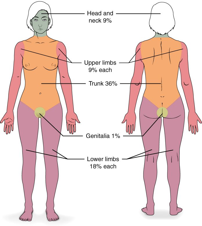
With labels hidden, select a box to reveal its label.
5How it works, step by step
- A large partial- and full-thickness burn destroys the stratum corneum and damages the capillaries of the dermis.Water evaporates freely from the wound, and damaged cells and mast cells release histamine and other chemical messengers.
- The chemical messengers reach capillaries under the burn and, in a large burn, throughout the body.The capillaries become leaky, and water, salts and proteins move from the plasma into the interstitial fluid.
- Fluid leaves the vessels by evaporation and leakage faster than it is replaced.Blood volume falls, so less blood returns to the heart.
- Less blood returns to the heart and less is pumped into the arteries.Blood pressure falls, the heart rate rises to compensate, and blood flow to the kidneys and urine output fall.
6Core concepts
7A common mistake
The wrong idea: A burn that does not hurt must be a mild burn.
What actually happens: A full-thickness burn destroys the whole dermis, including its nerve endings, so its center is painless, dry and leathery. It is the most serious kind of burn and cannot regrow epidermis on its own. The burns that hurt most are partial-thickness burns, whose nerve endings are irritated but still alive.
8Check yourself
Anything you miss goes into your review queue.
1. A 70 kg adult has partial- and full-thickness burns over 40% of his body surface. No fluid has been given yet. Predict the change in each variable over the first several hours, compared with before the burn.
| Variable | Change |
|---|---|
| Water lost by evaporation from the skin | — |
| Volume of interstitial fluid | — |
| Blood volume | — |
| Blood pressure | — |
| Heart rate | — |
| Urine output | — |
Show the answer
Across the integumentary, cardiovascular and urinary systems: a large burn removes the skin's barrier and makes capillaries leak body-wide. Water evaporates from the wound and plasma leaks into the tissues, so blood volume and blood pressure fall, heart rate rises to compensate, and urine output falls. That is why large burns need large volumes of fluid in the first day.
- Water lost by evaporation from the skin: up. The burned skin has lost its stratum corneum and lipid seal, so water evaporates freely from the wound surface.
- Volume of interstitial fluid: up. Heat damage and chemical messengers make capillaries leaky, so water, salts and proteins move from the plasma into the spaces between cells, and the tissue swells.
- Blood volume: down. Fluid leaves the vessels into the tissues and out through the burned surface faster than it is replaced.
- Blood pressure: down. With less blood volume, less blood returns to the heart and less is pumped into the arteries.
- Heart rate: up. The body responds to the falling blood pressure by speeding the heart.
- Urine output: down. Low blood pressure reduces blood flow to the kidneys, so they make less urine.
2. Using the adult rule of nines, what share of body surface is burned in a patient with partial-thickness burns over the whole front of the trunk and the whole left arm?
- 18%
- 27%
- 36%
- 45%
Show the answer
Front of the trunk = 18%; one whole upper limb = 9%. 18 + 9 = 27%.
- 18%: 18% is the front of the trunk alone; the arm adds another 9%.
- Correct: 27%: Correct. 18 + 9 = 27%.
- 36%: 36% is the whole trunk, front and back; only the front is burned.
- 45%: 45% would be the whole trunk plus one arm.
3. After a day at the beach, an adult has a red, painful sunburn with no blisters over the whole back of the trunk. He also spilled boiling water on his whole right arm, which is blistered. What share of body surface counts as burned for fluid planning?
- 27%
- 18%
- 9%
- 36%
Show the answer
Only partial- and full-thickness burns count. The sunburn is superficial (red, painful, no blisters) and leaves the barrier intact, so it is left out. The blistered arm is partial-thickness: 9%.
- 27%: 27% adds the superficial sunburn, which is not counted.
- 18%: 18% is the sunburned back alone, which does not count.
- Correct: 9%: Correct. Only the arm's 9% counts.
- 36%: 36% is the whole trunk; neither the burns nor the rule support it.
4. After a house fire, a patient's thigh has a white, leathery, dry area that he says does not hurt when touched, surrounded by a ring of red, blistered skin that is very painful. Which interpretation is correct?
- The painless center is a mild superficial burn
- The whole area is one partial-thickness burn
- The painless center is full-thickness; the painful ring is partial-thickness
- The ring is full-thickness, and the numb center is a burn that has already healed
Show the answer
A full-thickness burn destroys the whole dermis with its nerve endings, so its center is painless, white and leathery. The partial-thickness burn around it still has living nerve endings and capillaries, so it hurts and blisters. Painlessness here means the deepest damage.
- The painless center is a mild superficial burn: A superficial burn is red and painful, not white, leathery and numb.
- The whole area is one partial-thickness burn: Partial-thickness burns hurt and blister; a numb, leathery area has lost its nerve endings, which means full thickness.
- Correct: The painless center is full-thickness; the painful ring is partial-thickness: Correct. The numb center is the deepest part of the burn.
- The ring is full-thickness, and the numb center is a burn that has already healed: A red, blistered, painful ring has living nerve endings, which rules out full thickness; a burn does not heal from its center outward within hours.
5. A superficial partial-thickness burn destroys the whole epidermis over a patch of skin, yet it grows new epidermis in 2 to 3 weeks without a graft. Where do the new epidermal cells come from?
- Fibroblasts of the reticular layer, which change into new keratinocytes
- Fat cells of the hypodermis
- Melanocytes that survive the burn
- Epithelial cells lining surviving hair follicles and sweat gland ducts
Show the answer
Hair follicles and sweat gland ducts are lined by epithelium that reaches deep into the dermis, below a partial-thickness burn. Those cells divide and spread out across the wound bed, and the islands grow together into new epidermis. A full-thickness burn destroys them, so it cannot regrow epidermis from within.
- Fibroblasts of the reticular layer, which change into new keratinocytes: Fibroblasts make collagen and scar tissue; they do not become epithelial cells.
- Fat cells of the hypodermis: Fat cells lie below the dermis and do not form epidermis.
- Melanocytes that survive the burn: Melanocytes make pigment; they do not supply keratinocytes.
- Correct: Epithelial cells lining surviving hair follicles and sweat gland ducts: Correct. The epithelium of follicles and ducts regrows the surface.
6. A dog bite leaves a gaping wound with missing tissue on a man's calf. To lower the risk of infection, the doctor leaves it open to heal. Compared with a stitched surgical cut, how will it heal?
- By primary union, with a thin line scar within days
- By regeneration only, with no scar
- By secondary union, with granulation tissue and contraction
- By primary union, once the scab falls off and the edges meet
Show the answer
A wound with separated edges and lost tissue heals by secondary union. It fills from the bottom with granulation tissue, its edges are pulled together by wound contraction, and it closes over weeks, leaving a wider scar.
- By primary union, with a thin line scar within days: Primary union needs edges held close together, as in a stitched cut; this wound is left open.
- By regeneration only, with no scar: A wound through the dermis with lost tissue fills partly with collagen, so it scars.
- Correct: By secondary union, with granulation tissue and contraction: Correct. Open wounds heal by secondary union.
- By primary union, once the scab falls off and the edges meet: Whether a wound heals by primary or secondary union depends on whether its edges are together, not on the scab.
7. Melanoma makes up only about 1 in 100 skin cancers but causes most skin cancer deaths. What best explains this?
- Melanoma cells can spread early to lymph nodes and distant organs
- Melanoma grows only on skin that cannot be seen
- Melanoma cannot be removed by surgery
- Melanoma always starts in an old mole
Show the answer
Melanoma starts in melanocytes, which can break loose early, pass through the dermis into vessels and spread to lymph nodes and distant organs. Basal cell carcinoma rarely spreads, so it is common but rarely deadly.
- Correct: Melanoma cells can spread early to lymph nodes and distant organs: Correct. Early spread is what makes melanoma dangerous.
- Melanoma grows only on skin that cannot be seen: Melanoma can appear anywhere, including easily seen skin; that is why skin checks work.
- Melanoma cannot be removed by surgery: Thin melanomas removed early are usually cured.
- Melanoma always starts in an old mole: Most melanomas appear as new spots rather than in an existing mole.
8. A frail patient has lain on his back without moving for many hours. The skin over his heels is red and does not blanch when pressed. What is the main mechanism of the damage?
- Pressure squeezed his capillaries shut, starving cells of oxygen
- Heat from the mattress has burned the skin
- Bacteria from the sheets have infected the intact skin of his heels
- Too much sebum has plugged the follicles of the heel
Show the answer
Sustained pressure over a bony area, higher than the pressure inside the capillaries, flattens them. Blood stops flowing, the cells run out of oxygen and nutrients, and they begin to die by necrosis. Redness that does not blanch is the first sign of a pressure injury.
- Correct: Pressure squeezed his capillaries shut, starving cells of oxygen: Correct. Compressed capillaries starve the tissue.
- Heat from the mattress has burned the skin: A mattress is not hot enough to burn skin; the damage comes from pressure.
- Bacteria from the sheets have infected the intact skin of his heels: Infection can follow once skin breaks down, but the first damage is from lost blood flow.
- Too much sebum has plugged the follicles of the heel: The heel is thick skin with no hair and no sebaceous glands.
9Summary
Whether skin heals without a scar depends on what survives: basal cells for epidermal wounds, follicle and duct epithelium for partial-thickness burns, nothing for full-thickness burns. Wounds heal through a clot, inflammation, granulation tissue with new epidermis and wound contraction, then remodeling; closed edges heal by primary union, open wounds by secondary union, and a keloid is a scar that overgrows the wound. Burns are superficial (epidermis), partial-thickness (into the dermis, blistered, painful), full-thickness (through the dermis, leathery, painless) or fourth-degree (into muscle or bone). The rule of nines estimates the burned area in adults (head 9, each arm 9, trunk 18 front and 18 back, each leg 18, genital area 1), counting only partial- and full-thickness burns. Large burns lose fluid by evaporation and leaky capillaries, lowering blood volume and pressure. UV-driven mutations cause basal cell carcinoma, squamous cell carcinoma and melanoma, which spreads earliest. Pressure injuries, acne, stretch marks, calluses and corns, albinism, vitiligo and eczema each trace to one structure of the skin.