Skeletal muscle structure
1Why this matters
Nine-year-old Leo has trouble climbing stairs, so his doctor sends a tiny piece of his thigh muscle to the lab. Under the microscope, the pathologist checks each level of the muscle in turn: are the bundles and their wrappings normal, are the fibers the right size with their nuclei at the edge, and are the stripes crisp and evenly spaced? Reading that slide means knowing how a skeletal muscle is built, from the whole organ down to its protein filaments.
2What this builds on
3Quick check before you start
1. Which kind of connective tissue forms tendons?
- Dense regular connective tissue
- Loose areolar connective tissue
- Adipose tissue
Show the answer
Tendons are dense regular connective tissue: tightly packed collagen fibers all running in one direction, which resists pulling along that line.
- Correct: Dense regular connective tissue:
- Loose areolar connective tissue:
- Adipose tissue:
2. What does the smooth endoplasmic reticulum do in most cells?
- Makes proteins for export on its ribosomes
- Makes lipids and stores calcium ions
- Packages proteins into vesicles for secretion
Show the answer
Smooth ER has no ribosomes. It makes lipids and, in many cells, stores calcium ions. In muscle it is specialized for calcium storage.
- Makes proteins for export on its ribosomes:
- Correct: Makes lipids and stores calcium ions:
- Packages proteins into vesicles for secretion:
3. Skeletal muscle fibers look striped under a microscope. What is a skeletal muscle fiber?
- A protein thread in the extracellular matrix
- A bundle of many small cells
- One long cell with many nuclei
Show the answer
A muscle fiber is one whole cell. In the embryo many cells fuse to form it, so it has many nuclei.
- A protein thread in the extracellular matrix:
- A bundle of many small cells:
- Correct: One long cell with many nuclei:
4Anatomy
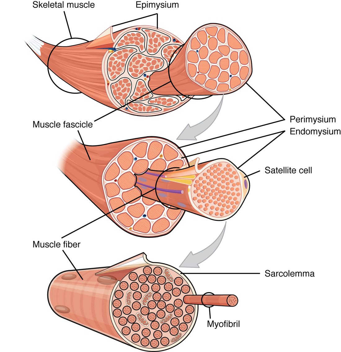
With labels hidden, select a box to reveal its label.
5How it works, step by step
- Myosin heads on the thick filaments pull the thin filaments toward the M line.Each sarcomere shortens as its Z discs move closer together.
- Thousands of sarcomeres, joined end to end in every myofibril, shorten at the same time.Each myofibril shortens, and the muscle fiber that holds it shortens.
- Each shortening fiber pulls on the endomysium around it.The pull passes to the perimysium around its fascicle and the epimysium around the whole muscle.
- The three wrappings merge into the tendon, whose collagen runs into the bone.The tendon pulls on the bone, and the bone moves at its joint.
6Core concepts
7A common mistake
The wrong idea: A myofibril and a muscle fiber are the same thing.
What actually happens: A muscle fiber is a whole cell, with a sarcolemma, many nuclei and mitochondria. A myofibril is a rod of protein filaments inside that cell, one of hundreds or thousands packed in its sarcoplasm. Myofibrils have no membrane and no nuclei of their own. Fibers are cells; myofibrils are inside cells; filaments are inside myofibrils.
8Check yourself
Anything you miss goes into your review queue.
1. Name the pinned connective tissue layer.
- Perimysium
- Epimysium
- Endomysium
- Sarcolemma
Show the answer
The perimysium wraps each fascicle, a bundle of muscle fibers. The larger blood vessels and nerves run into the muscle along it.
- Correct: Perimysium: Correct. The layer around each bundle of fibers is the perimysium (peri- = around).
- Epimysium: The epimysium wraps the whole muscle, outside all the fascicles.
- Endomysium: The endomysium is the thin layer around each single fiber, inside a fascicle.
- Sarcolemma: The sarcolemma is the plasma membrane of one fiber, not a connective tissue layer.
2. Put these levels of skeletal muscle structure in order, from largest to smallest.
- Whole muscle
- Fascicle
- Muscle fiber
- Myofibril
- Sarcomere
- Thick filament
Show the answer
A muscle is a bundle of fascicles; a fascicle is a bundle of fibers (cells); each fiber is packed with myofibrils; each myofibril is a chain of sarcomeres; and each sarcomere is built from thick and thin filaments.
- Correct order: 1. Whole muscle 2. Fascicle 3. Muscle fiber 4. Myofibril 5. Sarcomere 6. Thick filament
3. A researcher cuts across a sarcomere and sees only thin filaments in the cross-section. Where was the cut?
- Through the H zone
- Through the I band
- Through the M line
- Through the A band outside the H zone
Show the answer
The I band is the part of the sarcomere where thin filaments are not overlapped by thick ones, so a cut there shows thin filaments alone.
- Through the H zone: The H zone holds thick filaments only; the thin filaments do not reach that far in.
- Correct: Through the I band: Correct. The I band contains thin filaments only.
- Through the M line: The M line is at the center of the thick filaments, where proteins link them; a cut there shows thick filaments.
- Through the A band outside the H zone: Outside the H zone the A band has both kinds, each thick filament ringed by six thin ones.
4. A pathologist looks at a muscle biopsy cut across. Each individual fiber is surrounded by a thin layer of connective tissue that contains capillaries. Which layer is this?
- Epimysium
- Perimysium
- Deep fascia
- Endomysium
Show the answer
The endomysium surrounds each single muscle fiber and holds the capillaries that supply it with oxygen and fuel.
- Epimysium: The epimysium is a thick layer around the whole muscle, not around single fibers.
- Perimysium: The perimysium surrounds groups of fibers (fascicles), not each fiber.
- Deep fascia: Deep fascia lies outside whole muscles.
- Correct: Endomysium: Correct. A thin layer around each fiber, carrying capillaries, is the endomysium.
5. A skeletal muscle fiber is 80 µm thick, yet the myofibrils at its center start contracting almost as soon as those at its surface. What makes this possible?
- Calcium from the extracellular fluid diffuses quickly to the center
- Gap junctions pass the signal from myofibril to myofibril
- T tubules carry the electrical signal from the sarcolemma into the fiber's depths
- The nuclei at the center relay the signal
Show the answer
T tubules are infoldings of the sarcolemma that reach every myofibril. The electrical signal travels down them to the triads throughout the fiber, where the SR beside each myofibril holds the calcium.
- Calcium from the extracellular fluid diffuses quickly to the center: Calcium diffusing inward from the surface would reach the center far too slowly; the calcium used comes from the SR inside the fiber.
- Gap junctions pass the signal from myofibril to myofibril: Myofibrils are not cells, so there are no gap junctions between them.
- Correct: T tubules carry the electrical signal from the sarcolemma into the fiber's depths: Correct. T tubules bring the surface membrane, and its signal, to every triad.
- The nuclei at the center relay the signal: A skeletal muscle fiber's nuclei lie at its edge, just under the sarcolemma, and they do not relay signals.
6. In a resting muscle fiber, the myosin heads are loaded with energy, yet they do not bind actin. What prevents them?
- Tropomyosin covers the myosin-binding sites on actin
- The thick and thin filaments do not overlap at rest
- The myosin heads have no ATP bound
- Titin holds the myosin heads against the thick filament
Show the answer
At rest, tropomyosin lies over the myosin-binding sites on actin, held in place by troponin. Until calcium binds troponin, the heads cannot reach actin.
- Correct: Tropomyosin covers the myosin-binding sites on actin: Correct. Tropomyosin blocks the binding sites while calcium is low.
- The thick and thin filaments do not overlap at rest: The filaments do overlap at rest, throughout the A band outside the H zone.
- The myosin heads have no ATP bound: The heads carry the products of split ATP at rest; lack of ATP is not what holds them off.
- Titin holds the myosin heads against the thick filament: Titin centers the thick filament and gives elasticity; it does not restrain the heads.
7. A muscle fiber is 3 cm long and its sarcomeres are 2.5 µm long. About how many sarcomeres lie end to end along one of its myofibrils?
- 1,200
- 120,000
- 75,000
- 12,000
Show the answer
Convert to the same unit: 3 cm = 30,000 µm. Then divide: 30,000 µm ÷ 2.5 µm = 12,000 sarcomeres.
- 1,200: This result comes from converting 3 cm to 3,000 µm; 1 cm is 10,000 µm, not 1,000.
- 120,000: This comes from converting 3 cm to 300,000 µm; 1 cm is 10,000 µm, not 100,000.
- 75,000: This multiplies 30,000 µm by 2.5 instead of dividing the fiber's length by one sarcomere's length.
- Correct: 12,000: Correct. 30,000 µm ÷ 2.5 µm = 12,000.
9Summary
A skeletal muscle is bundles within bundles. The epimysium wraps the whole muscle, the perimysium wraps each fascicle, and the endomysium wraps each fiber; all three merge into the tendon, which carries the pull to bone. A muscle fiber is one cell: its plasma membrane is the sarcolemma and its cytoplasm the sarcoplasm, packed with myofibrils, mitochondria and glycogen. Each myofibril is a chain of sarcomeres, the functional units, running from Z disc to Z disc. Thick filaments of myosin form the dark A band, with the H zone and M line at its center; thin filaments of actin, anchored at the Z discs, reach into it and alone form the light I band. On the thin filament, tropomyosin covers the myosin-binding sites on actin, held there by troponin, which can bind calcium. The sarcoplasmic reticulum stores calcium around every myofibril, and T tubules, infoldings of the sarcolemma, carry the electrical signal deep into the fiber, where each one meets two terminal cisternae in a triad.