Muscles of the hip and lower limb
1Why this matters
Marcus, 22, breaks his tibia in a soccer tackle. Six hours after it is set, the front of his leg is tense and the pain is far worse than a fracture should cause. When the nurse gently points his toes, he cries out. His foot pulse is still there. The surgeon takes him straight back to the operating room to slit open the tough sheet of fascia around the muscles on the front of his leg.
2What this builds on
3Quick check before you start
1. Which bone marking is the bump you sit on?
- The ischial tuberosity
- The greater trochanter
- The anterior superior iliac spine
Show the answer
The ischial tuberosity, on the ischium, bears your weight when you sit. The greater trochanter is the bump on the upper outer femur, and the anterior superior iliac spine is the front point of the iliac crest.
- Correct: The ischial tuberosity:
- The greater trochanter:
- The anterior superior iliac spine:
2. Pointing the toes downward, as when you press a gas pedal, is which movement?
- Dorsiflexion
- Plantar flexion
- Eversion
Show the answer
Plantar flexion moves the sole downward, pointing the toes. Dorsiflexion lifts the toes toward the shin, and eversion turns the sole outward.
- Dorsiflexion:
- Correct: Plantar flexion:
- Eversion:
3. What is the deep fascia?
- A layer of fat under the skin
- Dense connective tissue that wraps muscles and holds them in groups
- The connective tissue around a single muscle fiber
Show the answer
Deep fascia is dense connective tissue beneath the subcutaneous layer that wraps muscles and binds them into groups. The fat under the skin is the subcutaneous tissue, and the endomysium surrounds single fibers.
- A layer of fat under the skin:
- Correct: Dense connective tissue that wraps muscles and holds them in groups:
- The connective tissue around a single muscle fiber:
4Anatomy
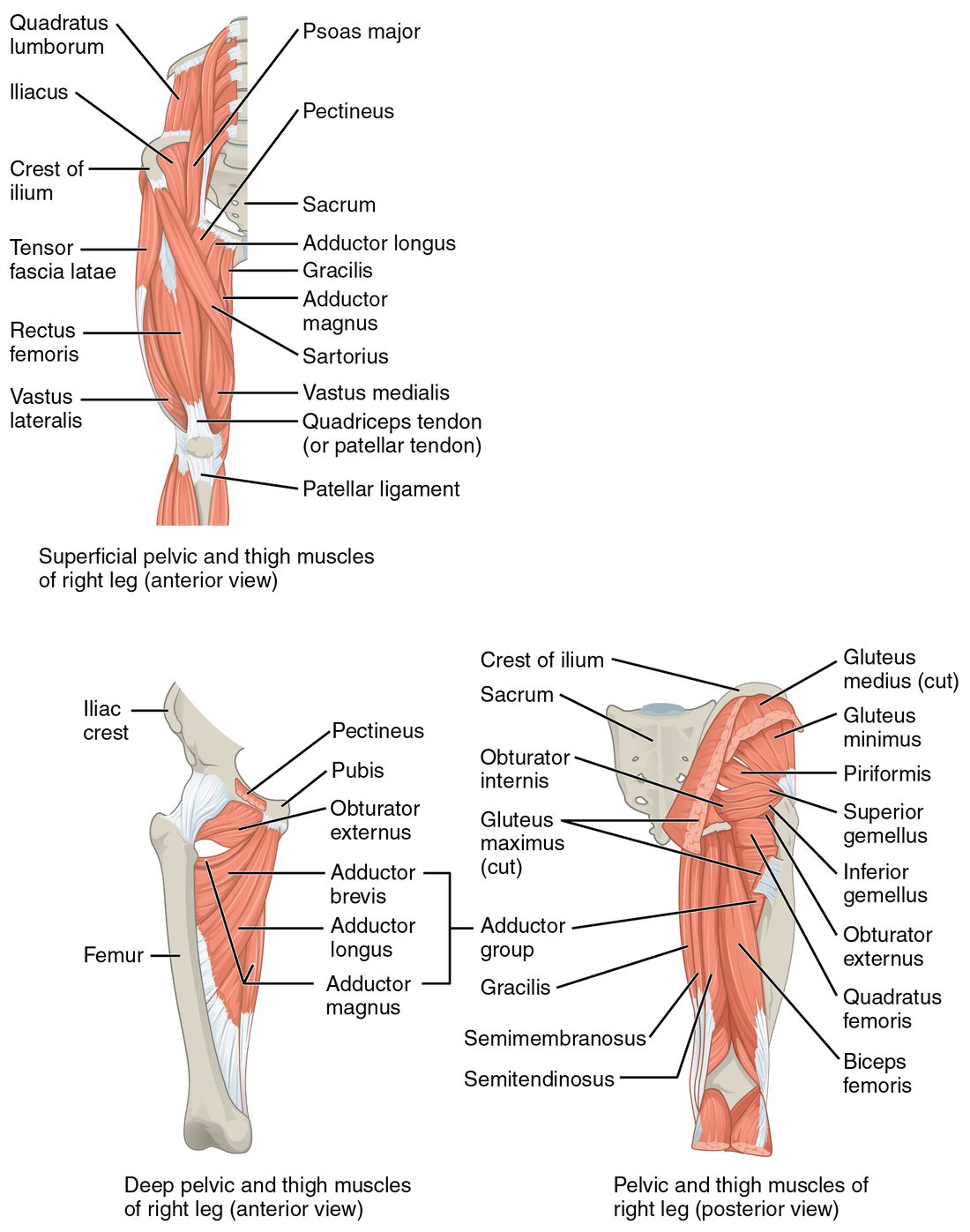
With labels hidden, select a box to reveal its label.
5How it works, step by step
- A tibia fracture bleeds and the injured muscles swell inside the anterior compartment of the leg.The volume inside the compartment grows.
- The deep fascia and bones around the compartment barely stretch.The pressure inside the compartment rises.
- The rising pressure squeezes the thin-walled veins shut.Blood cannot drain out, more fluid is trapped, and the pressure climbs further.
- The pressure rises high enough to stop blood flow through the capillaries.The muscles and nerves in the compartment are starved of oxygen, causing severe pain that worsens when those muscles are stretched.
- A surgeon slits the fascia along the compartment.The pressure falls and blood flow returns before the muscle dies.
6Core concepts
7A common mistake
The wrong idea: If the pulse in the foot is present, compartment syndrome is ruled out.
What actually happens: The pressure inside the main artery is far higher than the pressure that stops capillary flow in a compartment, so blood can still pulse through the artery while the muscles around it starve. A lost pulse is a late sign. Pain out of proportion to the injury and pain on passive stretch come first.
8Check yourself
Anything you miss goes into your review queue.
1. Name the pinned muscle running straight down the middle of the front of the thigh.
- Vastus intermedius
- Sartorius
- Rectus femoris
- Vastus lateralis
Show the answer
The rectus femoris runs straight down the middle of the front of the thigh from the ilium to the quadriceps tendon. It is the only part of the quadriceps femoris that crosses the hip.
- Vastus intermedius: The vastus intermedius lies deep, hidden under the rectus femoris.
- Sartorius: The sartorius crosses the thigh diagonally, from the anterior superior iliac spine to the medial tibia.
- Correct: Rectus femoris: Correct. The straight middle muscle is the rectus femoris.
- Vastus lateralis: The vastus lateralis is on the outer side of the thigh.
2. On the back of the thigh, name the pinned lateral hamstring.
- Semimembranosus
- Biceps femoris
- Semitendinosus
- Vastus lateralis
Show the answer
The biceps femoris is the lateral hamstring. Its long head starts on the ischial tuberosity and its short head on the linea aspera; it inserts on the head of the fibula.
- Semimembranosus: The semimembranosus is a medial hamstring.
- Correct: Biceps femoris: Correct. The lateral hamstring is the biceps femoris.
- Semitendinosus: The semitendinosus is a medial hamstring, with a long cord-like tendon.
- Vastus lateralis: The vastus lateralis is on the outer front of the thigh, part of the quadriceps femoris.
3. Six hours after a tibia fracture was set, a 22-year-old soccer player has severe pain in the front of his leg, far worse than expected, and it gets much worse when the nurse gently points his toes downward. His foot pulse can still be felt. What is the most likely explanation?
- Compartment syndrome of the anterior compartment of the leg
- Rupture of the calcaneal tendon
- A hamstring strain
- Normal pain after a fracture is set
Show the answer
Bleeding and swelling inside the anterior compartment, wrapped in deep fascia that cannot stretch, raise the pressure until blood cannot flow through the capillaries. Pain out of proportion to the injury, and pain when the compartment's muscles are passively stretched (pointing the toes stretches the dorsiflexors), are the early signs. A pulse is often still present.
- Correct: Compartment syndrome of the anterior compartment of the leg: Correct. This is compartment syndrome of the front of the leg.
- Rupture of the calcaneal tendon: A calcaneal tendon rupture causes weakness rising onto the toes and pain at the back of the ankle, not severe pain in the front of the leg.
- A hamstring strain: The hamstrings are on the back of the thigh, not the front of the leg.
- Normal pain after a fracture is set: Pain far worse than expected, and worse when the compartment's muscles are stretched, is not normal. A present pulse does not rule compartment syndrome out: the pressure in the large artery is far higher than the pressure in the compartment, so the pulse is often felt until late.
4. Which muscles belong to the quadriceps femoris? Select all that apply.
- Rectus femoris
- Vastus lateralis
- Sartorius
- Vastus medialis
- Vastus intermedius
- Biceps femoris
Show the answer
The quadriceps femoris is four muscles: the rectus femoris and the vastus lateralis, medialis and intermedius. All four insert through the quadriceps tendon, patella and patellar ligament on the tibial tuberosity.
- Correct: Rectus femoris: Yes. The rectus femoris is the one part that crosses the hip.
- Correct: Vastus lateralis: Yes. The vastus lateralis is on the outer thigh.
- Sartorius: No. The sartorius is also in the front of the thigh, but it inserts on the medial tibia and is not part of the quadriceps.
- Correct: Vastus medialis: Yes. The vastus medialis is on the inner thigh above the knee.
- Correct: Vastus intermedius: Yes. The vastus intermedius lies deep under the rectus femoris.
- Biceps femoris: No. The biceps femoris is a hamstring, on the back of the thigh.
5. When a patient stands on her right leg, the left side of her pelvis drops. Weakness of which muscle best explains this?
- Left gluteus maximus
- Right gluteus medius
- Right iliopsoas
- Left adductor longus
Show the answer
Standing on the right leg, the body's weight hangs to the left of the right hip. The right gluteus medius and minimus, abductors of the standing leg, pull the ilium down toward the greater trochanter and hold the pelvis level. When they are weak, the opposite side drops: a positive Trendelenburg sign.
- Left gluteus maximus: The gluteus maximus mainly extends the hip, and the muscle that levels the pelvis belongs to the standing leg, here the right.
- Correct: Right gluteus medius: Correct. The standing leg's gluteus medius holds the pelvis level.
- Right iliopsoas: The iliopsoas flexes the hip; it does not hold the pelvis level in standing.
- Left adductor longus: The adductor longus adducts the thigh and belongs to the lifted leg; it cannot hold up the pelvis on this side.
6. A nurse gives a routine vaccine to a 4-month-old infant. Which site is preferred?
- Deltoid site
- Dorsogluteal site
- Vastus lateralis site
- The gastrocnemius
Show the answer
An infant's thigh muscles are well developed while the gluteal and deltoid muscles are still small, so the middle third of the vastus lateralis on the outer thigh is the preferred site.
- Deltoid site: The deltoid is the usual vaccine site in adults, but in young infants it is too small.
- Dorsogluteal site: The dorsogluteal site is avoided at every age: a large nerve lies close by and the fat layer is thick.
- Correct: Vastus lateralis site: Correct. The vastus lateralis is preferred in infants.
- The gastrocnemius: The calf is not a standard injection site.
7. This traces the hamstrings from origin to insertion. One step is wrong. Find it.
- All three originate on the ischial tuberosity
- They run down the back of the thigh, behind the hip joint
- They cross behind the knee joint
- They insert on the tibial tuberosity
Show the answer
The hamstrings insert behind the knee's axis, on the head of the fibula (biceps femoris) and the medial tibia (semitendinosus and semimembranosus). The tibial tuberosity, on the front of the tibia, is where the quadriceps femoris inserts.
- All three originate on the ischial tuberosity: Correct step. The ischial tuberosity is the hamstrings' shared origin.
- They run down the back of the thigh, behind the hip joint: Correct step. Running behind the hip, they extend it.
- They cross behind the knee joint: Correct step. Crossing behind the knee, they flex it.
- Correct: They insert on the tibial tuberosity: This is the wrong step. They insert on the fibula and medial tibia, not the tibial tuberosity.
8. Walking on a sloping trail, you turn the soles of your feet outward to keep them flat. Which muscles are producing this eversion?
- Tibialis anterior and tibialis posterior
- Gastrocnemius and soleus
- Fibularis longus and fibularis brevis
- Extensor digitorum longus and sartorius
Show the answer
The fibularis longus and brevis, in the lateral group of the leg, pass behind the lateral malleolus to the foot and turn the sole outward.
- Tibialis anterior and tibialis posterior: Both tibialis muscles invert the foot, turning the sole inward.
- Gastrocnemius and soleus: The gastrocnemius and soleus plantar flex the foot through the calcaneal tendon.
- Correct: Fibularis longus and fibularis brevis: Correct. The fibularis muscles evert the foot.
- Extensor digitorum longus and sartorius: The extensor digitorum longus mainly extends the toes, and the sartorius acts at the hip and knee, not the foot.
9Summary
The iliopsoas flexes the hip and the gluteus maximus extends it; the gluteus medius and minimus abduct the thigh and hold the pelvis level. The quadriceps femoris extends the knee through the quadriceps tendon, patella and patellar ligament, and the rectus femoris also flexes the hip. The adductors adduct the thigh; the hamstrings flex the knee and extend the hip. In the leg, the front group dorsiflexes, the side group everts and the back group plantar flexes the foot through the calcaneal tendon. Deep fascia divides each limb segment into compartments of muscles with one shared action; trapped pressure in one causes compartment syndrome. The deltoid, ventrogluteal and vastus lateralis injection sites are chosen for thick muscle, clear landmarks and no large nerves nearby.