Classifying joints
1Why this matters
Ms. Reyes is 32 weeks pregnant and has sharp pain at the front of her pelvis every time she climbs stairs or stands on one leg. An ultrasound shows that the gap between her two pubic bones, usually 4 to 5 mm, has widened to 12 mm. Nothing is broken. In pregnancy the tissue of this joint loosens and the gap normally widens by a few millimeters, but in Ms. Reyes it has widened more than usual.
2What this builds on
3Quick check before you start
1. Which kind of cartilage forms the intervertebral discs and the menisci of the knee?
- Hyaline cartilage
- Fibrocartilage
- Elastic cartilage
Show the answer
Fibrocartilage is packed with thick collagen bundles, so it resists both squeezing and pulling. It forms the intervertebral discs and the knee menisci. Hyaline cartilage covers bone ends, and elastic cartilage forms the ear flap.
- Hyaline cartilage:
- Correct: Fibrocartilage:
- Elastic cartilage:
2. What does a ligament join?
- Muscle to bone
- Bone to bone
- Skin to muscle
Show the answer
A ligament is a cord of dense regular connective tissue that joins bone to bone. A tendon joins muscle to bone.
- Muscle to bone:
- Correct: Bone to bone:
- Skin to muscle:
3. What does a synovial membrane do?
- Lines the cavities of movable joints and releases a slippery fluid
- Covers the outer surface of a bone
- Lines the body cavities and covers the organs in them
Show the answer
A synovial membrane is a connective tissue membrane that lines the spaces inside movable joints and releases a thick, slippery fluid. The periosteum covers bone, and serous membranes line body cavities.
- Correct: Lines the cavities of movable joints and releases a slippery fluid:
- Covers the outer surface of a bone:
- Lines the body cavities and covers the organs in them:
4Anatomy
With labels hidden, select a box to reveal its label.
5How it works, step by step
- During pregnancy, hormones loosen the collagen in the fibrocartilage pad and the ligaments of the pubic symphysis.The pad and ligaments stretch more when a load pulls or shears across them.
- The joint's tissue stretches more under each load.The gap between the two pubic bones widens, and the bones shift more each time weight moves from one leg to the other.
- The bones shift farther than usual against each other.The joint surfaces shear and stretch pain-sensitive tissue, so standing on one leg or climbing stairs hurts at the front of the pelvis.
- After delivery, the hormone levels fall and the collagen tightens again.The tissue stretches less, the gap narrows back toward 4 to 5 mm, and the joint returns to being only slightly movable.
6Core concepts
7A common mistake
The wrong idea: A joint is a place where bones move.
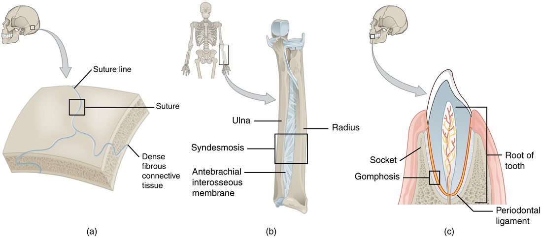
What actually happens: A joint is any place where a bone meets another bone, a cartilage or a tooth. Many joints barely move or do not move at all: the sutures of an adult skull and the joint between a tooth and its socket are joints, and they are classified as synarthroses (immobile joints). How much a joint moves depends on the tissue between the bones.
8Check yourself
Anything you miss goes into your review queue.
1. Which statement defines a joint (articulation)?
- Any place where a bone meets another bone, a cartilage or a tooth
- A place where two bones move freely against each other
- A cord of dense connective tissue that joins two bones
- The smooth cartilage that covers the end of a bone
Show the answer
A joint is defined by two structures meeting and being held together, not by movement. Many joints barely move.
- Correct: Any place where a bone meets another bone, a cartilage or a tooth: Correct. Any meeting point of bone with bone, cartilage or tooth is a joint.
- A place where two bones move freely against each other: This describes only freely movable joints. Skull sutures are joints too, and they do not move in an adult.
- A cord of dense connective tissue that joins two bones: This describes a ligament, which can help hold a joint together but is not the joint itself.
- The smooth cartilage that covers the end of a bone: This describes the cartilage on a bone end, one part found in some joints, not the joint itself.
2. Name the pinned tissue that holds the tooth in its socket.
- Interosseous membrane
- Periodontal ligament
- Articular cartilage
- Periosteum
Show the answer
The periodontal ligament is the thin layer of collagen fibers running from the tooth root to the socket wall. It forms the gomphosis.
- Interosseous membrane: An interosseous membrane is the broad sheet of fibers between two long bones, such as the radius and ulna.
- Correct: Periodontal ligament: Correct. The fibers around the tooth root are the periodontal ligament.
- Articular cartilage: Articular cartilage is hyaline cartilage covering bone ends, not fibers around a tooth root.
- Periosteum: The periosteum is the membrane covering the outer surface of a bone, not the fibers tying a tooth to its socket.
3. Which of these are fibrous joints? Select all that apply.
- Coronal suture
- Tooth in its socket
- Radius and ulna joined by an interosseous membrane
- Pubic symphysis
- Epiphyseal plate
Show the answer
Sutures, gomphoses and syndesmoses are the three fibrous joints. The pubic symphysis and the epiphyseal plate are cartilaginous.
- Correct: Coronal suture: Yes. A suture is a fibrous joint between skull bones.
- Correct: Tooth in its socket: Yes. A tooth in its socket is a gomphosis, a fibrous joint.
- Correct: Radius and ulna joined by an interosseous membrane: Yes. Bones joined by an interosseous membrane form a syndesmosis, a fibrous joint.
- Pubic symphysis: No. The pubic symphysis joins bones with fibrocartilage, so it is cartilaginous.
- Epiphyseal plate: No. The epiphyseal plate is hyaline cartilage, a synchondrosis.
4. A joint is made of a pad of fibrocartilage between two bones and allows a little bending. Which pair of labels fits it?
- Symphysis; amphiarthrosis
- Synchondrosis; synarthrosis
- Suture; synarthrosis
- Syndesmosis; diarthrosis
Show the answer
A fibrocartilage pad makes a symphysis, and slight movement makes it an amphiarthrosis. The intervertebral discs and the pubic symphysis fit this pair.
- Correct: Symphysis; amphiarthrosis: Correct. Fibrocartilage pad plus slight movement is a symphysis and an amphiarthrosis.
- Synchondrosis; synarthrosis: A synchondrosis uses hyaline cartilage and barely moves. This joint uses fibrocartilage and bends.
- Suture; synarthrosis: A suture is a fibrous joint between skull bones with no cartilage pad.
- Syndesmosis; diarthrosis: A syndesmosis is fibrous, not cartilaginous, and it is not freely movable.
5. Fill the gap. Suture : synarthrosis :: intervertebral disc : ____
- Amphiarthrosis
- Diarthrosis
- Synostosis
- Syndesmosis
Show the answer
A suture is an immobile joint, a synarthrosis. An intervertebral disc allows slight movement, so it is an amphiarthrosis.
- Correct: Amphiarthrosis: Correct. The disc is slightly movable.
- Diarthrosis: A diarthrosis is freely movable, like a synovial joint. One disc allows only slight movement.
- Synostosis: A synostosis is a joint that has turned to bone. A healthy disc is fibrocartilage.
- Syndesmosis: A syndesmosis is a structural subtype (a fibrous joint), not a functional class, and a disc is cartilaginous.
6. Which statement about the two classification schemes is accurate?
- The joint between the sacrum and hip bone moves freely
- Synchondroses are slightly movable amphiarthroses
- Fibrous joints include immobile and slightly movable ones
- The two schemes are two names for one grouping
Show the answer
Sutures and gomphoses are immobile fibrous joints, while syndesmoses are slightly movable fibrous joints. One structural class spans two functional classes.
- The joint between the sacrum and hip bone moves freely: The joint between the sacrum and hip bone is synovial but held so tightly by ligaments that it barely moves, so it is close to an amphiarthrosis.
- Synchondroses are slightly movable amphiarthroses: Synchondroses such as the epiphyseal plate are nearly immobile, so they are synarthroses, not amphiarthroses.
- Correct: Fibrous joints include immobile and slightly movable ones: Correct. Fibrous joints span synarthroses and amphiarthroses.
- The two schemes are two names for one grouping: The two schemes are separate. Structure predicts function most of the time, but that joint and the synchondroses show the groupings differ.
9Summary
A joint (articulation) is where a bone meets another bone, a cartilage or a tooth. Structurally, fibrous joints join bones with dense connective tissue: sutures in the skull, syndesmoses with a ligament or interosseous membrane, and gomphoses where the periodontal ligament holds a tooth. Cartilaginous joints join bones with hyaline cartilage (synchondrosis) or fibrocartilage (symphysis). Synovial joints separate the bone ends with a fluid-filled space. Functionally, a synarthrosis is immobile, an amphiarthrosis slightly movable and a diarthrosis freely movable. A synostosis is a joint that has turned to bone. The more a joint can move, the less stable it is.