Bone formation and growth
1Why this matters
Leo, 11, lands badly off a trampoline and breaks his leg just above the knee. The X-ray shows the break running through a band of cartilage near the end of his thigh bone. His surgeon lines the pieces up with great care and warns that, even after the bone heals, that leg may grow more slowly than the other. That band is where Leo's bone is still getting longer. An adult has no such band, and no longer grows in height.
2What this builds on
3Quick check before you start
1. What are the cells of cartilage called, and how do they get their nutrients?
- Chondrocytes, by diffusion through the matrix
- Osteocytes, through canaliculi
- Fibroblasts, from capillaries in the matrix
Show the answer
Chondrocytes sit in lacunae in cartilage matrix. Cartilage has no blood vessels, so nutrients reach them by diffusing through the matrix from the perichondrium.
- Correct: Chondrocytes, by diffusion through the matrix:
- Osteocytes, through canaliculi:
- Fibroblasts, from capillaries in the matrix:
2. Which part of a long bone is its widened end?
- The diaphysis
- The epiphysis
- The medullary cavity
Show the answer
Each widened end is an epiphysis; the shaft is the diaphysis, and the flared zone between them is the metaphysis.
- The diaphysis:
- Correct: The epiphysis:
- The medullary cavity:
3. Which bone cell lays down new bone matrix?
- Osteoclast
- Osteocyte
- Osteoblast
Show the answer
Osteoblasts secrete osteoid, which then mineralizes. Osteoclasts resorb bone, and osteocytes maintain it.
- Osteoclast:
- Osteocyte:
- Correct: Osteoblast:
4Anatomy
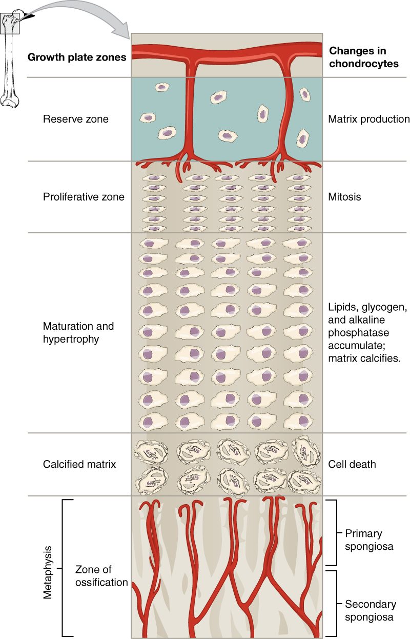
With labels hidden, select a box to reveal its label.
5How it works, step by step
- Chondrocytes in the proliferative zone divide and stack up, adding cells and matrix.The plate thickens on its epiphysis side, pushing the epiphysis away from the diaphysis.
- Older chondrocytes, pushed down the stack, stop dividing and swell.The matrix around the enlarged cells calcifies.
- Calcified matrix blocks diffusion of nutrients to the chondrocytes.Many of the chondrocytes die, leaving columns of calcified cartilage with empty spaces.
- Blood vessels and osteogenic cells from the diaphysis invade the spaces.Osteoblasts lay bone on the calcified columns, so the diaphysis grows longer while the plate keeps the same thickness.
6Core concepts
7A common mistake
The wrong idea: During development, cartilage hardens and turns into bone.
What actually happens: Cartilage does not harden into bone. In endochondral ossification, the cartilage model's matrix calcifies, and osteoblasts that arrive with invading blood vessels lay new bone on the leftover scaffold, which osteoclasts later clear away. Bone replaces cartilage. Calcified cartilage is still cartilage. Many of the enlarged chondrocytes die; studies that follow labeled cells in mice show that others survive and become bone cells.
8Check yourself
Anything you miss goes into your review queue.
1. An embryo's flat skull roof bones are forming. What are they forming from?
- Hyaline cartilage models
- Fibrocartilage
- Sheets of mesenchyme
- Epiphyseal plates
Show the answer
The flat bones of the skull roof form by intramembranous ossification: osteoblasts differentiate from mesenchymal cells within a sheet of mesenchyme and lay down bone directly, with no cartilage stage.
- Hyaline cartilage models: Cartilage models are the starting point for endochondral ossification, which forms the limb bones, vertebrae and skull base.
- Fibrocartilage: Fibrocartilage is not a template for bone formation.
- Correct: Sheets of mesenchyme: Correct. Skull roof bones form directly in mesenchyme.
- Epiphyseal plates: Epiphyseal plates are found in growing long bones, not in flat skull bones.
2. A gene change in achondroplasia slows chondrocyte division in the proliferative zone. Affected people have short arms and legs, while the dome of the skull grows to full size, often larger than average. Why is the skull dome spared?
- It forms by intramembranous ossification, with no epiphyseal plates
- Its epiphyseal plates close before birth, ending growth early
- Skull bones have no osteoblasts
- Its cartilage model is replaced by bone faster than in the limbs
Show the answer
The flat bones of the skull dome form directly in mesenchyme and grow at their edges, without cartilage models or epiphyseal plates. A defect in plate chondrocytes shortens the long bones, which lengthen at the plates, and spares bones that never use them.
- Correct: It forms by intramembranous ossification, with no epiphyseal plates: Correct. No cartilage, no plates, so a plate defect cannot slow them.
- Its epiphyseal plates close before birth, ending growth early: The skull dome never has epiphyseal plates at all; they are features of long bones.
- Skull bones have no osteoblasts: All bone, including skull bone, is laid down by osteoblasts.
- Its cartilage model is replaced by bone faster than in the limbs: The skull dome has no cartilage model to replace; that is the point of intramembranous ossification.
3. A child takes a drug for several months that blocks cell division in the proliferative zone of her epiphyseal plates. Predict the change in each variable, compared with before the drug.
| Variable | Change |
|---|---|
| Number of new chondrocytes made in the plate | — |
| Rate of growth in length | — |
| Appositional growth at the periosteum | — |
| Thickness of the epiphyseal plate | — |
Show the answer
Growth in length starts with cell division in the proliferative zone. Block it and less cartilage is made, lengthening slows, and the plate thins because replacement by bone continues. Growth in width uses a different mechanism and continues.
- Number of new chondrocytes made in the plate: down. The proliferative zone is where plate chondrocytes divide; blocking division stops new cells being made.
- Rate of growth in length: down. Fewer new cells means less cartilage added to push the epiphysis away, so there is less cartilage for bone to replace and the diaphysis lengthens more slowly.
- Appositional growth at the periosteum: no change. Width growth depends on osteoblasts under the periosteum, which the drug does not target.
- Thickness of the epiphyseal plate: down. Bone keeps replacing cartilage on the diaphysis side while little new cartilage is added on the epiphysis side, so the plate thins.
4. Put the steps of endochondral ossification of a long bone in order.
- Chondrocytes build a hyaline cartilage model
- The middle of the model calcifies as a bone collar forms around it
- A nutrient artery invades and a primary ossification center forms
- Osteoclasts open the medullary cavity
- Secondary ossification centers form in the epiphyses
Show the answer
A cartilage model forms first. Its middle calcifies and many of its chondrocytes die, while the perichondrium becomes periosteum and lays a bone collar. A nutrient artery brings in bone-forming cells, creating the primary ossification center; osteoclasts then hollow out the medullary cavity. Around birth and in childhood, the epiphyses ossify from secondary centers.
- Correct order: 1. Chondrocytes build a hyaline cartilage model 2. The middle of the model calcifies as a bone collar forms around it 3. A nutrient artery invades and a primary ossification center forms 4. Osteoclasts open the medullary cavity 5. Secondary ossification centers form in the epiphyses
5. A 24-year-old man hopes a supplement will make him taller. His knee X-ray shows a thin line of dense bone at the ends of his long bones where the growth plates once were. What can you tell him?
- His bones can still grow in length slowly
- His bones can grow in length if the line is stimulated
- His bones can no longer grow in length
- His bones can no longer grow in width
Show the answer
Those thin lines are epiphyseal lines. His plates have closed and been replaced by bone, so there is no cartilage left to grow interstitially. No supplement can add length. His bones can still thicken slowly by appositional growth.
- His bones can still grow in length slowly: Growth in length needs an open plate of cartilage. The lines show his plates have closed.
- His bones can grow in length if the line is stimulated: An epiphyseal line is bone, and bone cannot grow from within, so there is nothing to stimulate.
- Correct: His bones can no longer grow in length: Correct. Closed plates mean no more growth in length.
- His bones can no longer grow in width: Appositional growth at the periosteum continues through life, so width can still increase slowly.
6. During endochondral ossification, what happens to the cartilage of the model?
- It is converted into fibrocartilage
- It hardens into bone as minerals are added
- It stays inside the bone as a core
- It is broken down and replaced by bone
Show the answer
Cartilage does not harden into bone. Its matrix calcifies, many of its chondrocytes die, and invading osteoblasts lay bone on the leftover calcified columns, which osteoclasts later clear away. Bone replaces the cartilage.
- It is converted into fibrocartilage: The model is hyaline cartilage, and bone replaces it; it does not turn into another kind of cartilage.
- It hardens into bone as minerals are added: Calcified cartilage is still cartilage; it only serves as a temporary scaffold for new bone.
- It stays inside the bone as a core: Only the articular cartilage and, until growth ends, the epiphyseal plate remain. The rest is removed.
- Correct: It is broken down and replaced by bone: Correct. The model is a template that bone replaces.
7. As a child's long bone shaft gets wider, its wall stays about the same thickness relative to the bone. Which pair of events explains this?
- Osteoblasts add bone at the periosteum while osteoclasts remove it at the endosteum
- Chondrocytes divide in the shaft wall while osteoblasts add bone at the endosteum
- Osteoclasts remove bone at the periosteum while osteoblasts add bone at the endosteum
- The epiphyseal plate adds bone to the outside of the shaft
Show the answer
Appositional growth adds new bone under the periosteum, on the outside. Osteoclasts on the endosteum remove bone from the inside, widening the medullary cavity. The bone widens without its wall becoming ever thicker and heavier.
- Correct: Osteoblasts add bone at the periosteum while osteoclasts remove it at the endosteum: Correct. Build outside, remove inside.
- Chondrocytes divide in the shaft wall while osteoblasts add bone at the endosteum: There is no cartilage in the shaft wall to divide, and adding bone inside would narrow the cavity.
- Osteoclasts remove bone at the periosteum while osteoblasts add bone at the endosteum: That reversal would make the bone narrower, not wider.
- The epiphyseal plate adds bone to the outside of the shaft: The epiphyseal plate adds length at the ends; it does not add bone to the shaft's surface.
9Summary
Ossification is bone formation by osteoblasts, starting at ossification centers. In intramembranous ossification, bone forms directly in a sheet of mesenchyme: the skull roof, most bones of the face and the collarbones. In endochondral ossification, which forms most bones, a hyaline cartilage model is replaced by bone: its middle calcifies, a bone collar forms, a nutrient artery brings in bone-forming cells to make the primary ossification center in the diaphysis, osteoclasts open the medullary cavity, and secondary ossification centers form in the epiphyses. Cartilage remains as articular cartilage and as the epiphyseal plate, whose zones (reserve zone, proliferative zone, zone of maturation and hypertrophy, zone of calcified matrix) add cartilage that bone then replaces. That interstitial growth of cartilage lengthens the bone until the plates close in the late teens, leaving epiphyseal lines. Bones widen by appositional growth: osteoblasts add bone under the periosteum while osteoclasts remove it inside.